

 PDF
PDF ePub
ePub Citation
 Print
Print


I. Introduction
Nowadays, dental composite resins are used increasingly by practitioners for esthetic qualities. As presently posterior composites are suited to be bonded Class I and Class II cavity preparations.
The mechanism of adhesive bonding is based on acid etching both enamel and dentin of the tooth cavity surface. Although adhesion to phosphoric acid-etched enamel is reliable and long-lasting, adhesion to dentin has been more challenging because of the complex mineral and organic components of dentin. The bonding mechanism of adhesive resins to dentin proposed by Nakabayashi1) was described as micromechanical bonding due to the impregnation and polymerization of monomers into the exposed collagen of demineralized dentin surface, creating a hybrid layer. The major role of the adhesive resin is the stabilization of the formed hybrid layer and the formation of resin tag in the unplugged dentinal tubules2).
In the adhesive dentistry, total-etch technique has led to major improvements. However, the achievement of reliable bond to dental hard tissue without a separate acid etching step represented a major challenge in the past. Recently, self-etching adhesives were introduced. These systems use hydrophilic, acid monomers which are able to demineralize and penetrate enamel and dentin3). Unlike total-etch adhesives, these systems does not completely resolve or remove the smear layer. Kaaden et al.3) studied that the filled bonding agent Clearfil SE bond resulted in bond strengths higher than those of the unfilled adhesive systems. Perdigao and Lopes4) suggested that self-etching adhesives should provide optimum bonding besides a simplification of the bonding procedure and a potential decrease in technique sensitivity.
Most of the cavity preparations clinically show not only areas of exposed enamel and superficial dentin but also deep dentinal areas. Then variable location of exposed dentin such as cavity wall and cavity floor are appeared in the cut cavity. Thus a layer of cured adhesive of variable thickness is present on the cavity surface inevitably5). Adhesive thickness in the cavity wall is variable along the cavity depth by the gravity. In clinical situation, pooling of adhesive was apparent in the internal angle of the cavity and then decreased in thickness toward cavity margin. Peter et al.6) suggested that pooling of dentin bonding agents at the internal angle of the cavity arises because of the difference in viscosity between primers and unfilled adhesives. Perdigao et al.7,8) noticed that air thinning had a tendency to cause pooling of the adhesive into irregularities on the dentin surface and at the internal angle of the cavity. During the application of the adhesive, the manufacturer recommends that it should be applied in a uniform coating and then excessive air thinning of the adhesive should be avoided9).
Moreover, the variable adhesive thickness on the different area of the cavity would affect on the bond strength. Zheng et al.10) suggested that the effect of the thickness of the adhesive layer on bond strength is material-dependent. In their study, the increase in bond strengths of Clearfil Liner Bond 2V was directly proportional to the thickness of the bonding layer and the bond strengths of Single Bond decreased significantly with increase adhesive resin thickness. They emphasized that care should be taken to avoid excess adhesive resin at line angle in cavities bonded with single bottle system that contain water and ethanol10).
Recently, fillers have been added to single-bottle adhesive systems to reinforce the hybrid layer and increase bond strength. Increased filler loading increases viscosity of the bonding system and may reduce its flow11). In general, the variability of film thickness on the different site of the cavity is dependent on the type of bonding agents with or without filler because of the difference of their viscosity and flow. Grossman and Setzer5) showed that the bonding layer was thinnest at the cavity margin and thickest at the internal angle of the cavity for both type of the bonding agents. In addition, they described that layer thickness for filled adhesive, Optibond showed a progressive increase down the cavity wall to the internal angle while unfilled adhesive, Scotchbond Multi-purpose showed great film thickness variability5). Therefore, the variability of the adhesive thickness using of the different type of the bonding agents, filled adhesive and unfilled adhesive, should be evaluated on the prepared cavity.
The purposes of this study were to examine the variability of adhesive thickness on the different site of the cavity wall when used total-etch system without filler and simplified self-etch system with filler and to evaluate the relationship between variable adhesive thickness and microtensile bond strength to the cavity wall.
II. Materials and Methods
This study was performed in two parts. One part was to measure adhesive thickness on the cavity wall and the other part was to measure microtensile bond strength.
Part I. Adhesive thickness
Preparation of specimens
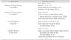
Six intact, non-carious, non-restored, human molars were used in this study. A class I cavity was prepared to expose all dentinal wall (mesiodistal width; 6 mm, buccolingual width; 4 mm, depth; 6 mm). After cavity preparation, three teeth were bonded with a filled adhesive (Clearfil™ SE bond; Kuraray, Medical Inc., Okayama, Japan) and the other three teeth were bonded with unfilled adhesives (Scotchbond™ Multi Purpose; 3M ESPE, St Paul, MN, USA) by Manufacture's recommendation. Rhodamine B was added in these adhesives. And then a hybrid light-activated resin composite (Z100; 3M ESPE, St. Paul, MN, USA) was filled in six cavities. The compositions of dentin bonding systems used in the study are described in Table 1.
The bonded teeth were then stored in distilled water at 37℃ for 24 hour prior to sectioning. Each tooth was sectioned bucco-lingually into a series of 1.0 mm thick slabs using a high-speed precision cut-off machine (Accutom-50; Struers, Ballerup, Denmark) with water coolant. Twelve specimens were prepared in each group (Table 2).
Fluorescence microscopy
These specimens were observed by fluorescence microscope (Axioskop; ZEISS, Oberkochen, Germany). Morphology and thickness of adhesive layer were founded through phase of diffusion with rhodamine B mixed adhesive resin. Bonding agent thickness was measured at three points along the cavity wall, edge of cavity margin (rim), halfway down each cavity wall (hlf), internal angle of the cavity (ang).
Statistical analysis
Statistical analysis of the collected data was performed by SPSS™ version 12.0 (SPSS Inc., Chicago, IL, USA). After calculating the means and standard deviations of the adhesive thickness for the specific points in each group, Student's t-test was used to compare adhesive thickness between two bonding systems. And one-way ANOVA and Scheffe's test for post-hoc comparison was performed to evaluate the difference of adhesive thickness among three points in each group. Statistical significance was defined as P < 0.05.
Part II. Microtensile bond strength
Preparation of specimens
Adhesive thickness at each point was reproduced by multiple coating of adhesive. Adhesive thickness at the rim and the hlf were same as one coat adhesive layer thickness in both Scotchbond™ Multi Purpose (SM group) and Clearfil™ SE bond (SE group), while adhesive thickness at the ang was same as seven coats in SM group and six coats in SE group.
Eight intact, non-carious, non-restored, human molars were used. A class I cavity was prepared to expose all dentinal wall. The teeth were sectioned longitudinally in mesiodistal direction through the prepared cavity using a diamond disc attached low-speed handpiece with air-water cooling. Sectioned sixteen teeth were classified into four groups.
The four groups were etched and bonded in manner described in Table 3. After dentin bonding procedure, a hybrid light-activated resin composite (Z100; 3M ESPE, St. Paul, MN, USA) was built up free hand in three increments to an approximate height of 6 mm. Each increment was 2 mm and light-cured for 20 s.
The teeth were stored in water at 37℃ for 24 hour. The 1 mm × 1 mm sticks were then sectioned mesiodistally and occlusogingivally using a high-speed precision cut-off machine under water coolant. Twenty specimens were prepared in each group.
Microtensile bond strength
The stick was fixed to the test bed using cyanoacrylate adhesive, Zapit (DVA Inc., Corona, CA, USA). The stick was pulled to failure under tension using a Micro Tensile Tester (Bisco inc., Shaumburg, IL, USA) at a crosshead speed of 1 mm/min. And then the microtensile bond strength was recorded in mpa.
Statistical analysis
Statistical analysis of the collected data was performed by SPSS™ version 12.0. After calculating the means and standard deviations of the microtensile bond strength for each group, Student's t-test was used to compare of microtensile bond strength for each group. Statistical significance was defined as P < 0.05.
III. Results
Part I. Adhesive thickness
Morphology of adhesive layer
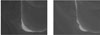
In fluorescence microscopy observation, adhesive layer was pooled only at the internal angle of the cavity for both bonding agents (Figure 1). And for both bonding agents the bonding layer was thinnest at the cavity margin and thickest at the internal angle of the cavity.
Clearfil™ SE bond formed uniform layer over the cavity wall compared to Scotchbond™ Multi Purpose.
Adhesive layer thickness
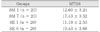
Table 4 shows the results of adhesive thickness at the specific area in two dentin bonding systems.
In Scotchbond™ Multi Purpose, adhesive thickness of ang was significantly thicker than that of rim and hlf (P < 0.05). Adhesive thickness of ang was also significantly thicker than that of rim and hlf when Clearfil™ SE bond was used (P < 0.05).
In rim and hlf, adhesive layer thickness of Clearfil SE bond was significantly thicker than that of Scotchbond™ Multi Purpose (P < 0.05). In ang , however, adhesive layer thickness of Scotchbond™ Multi Purpose was significantly thicker than that of Clearfil™ SE bond (P < 0.05).
Part II. Microtensile bond strength
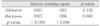
Table 5 and 6 show the results of microtensile bond strength for each group. Microtensile bond strength was highest in the SE 6 group (19.43 ± 3.68).
In Scotchbond™ Multi Purpose, seven coats group was significantly higher microtensile bond strength than one coat group (P < 0.05). In Clearfil™ SE bond, six coats group was also significantly higher microtensile bond strength than one coat group (P < 0.05). For both bonding agents, microtensile bond strength was increased as adhesive thickness was increased.
In one coat groups, microtensile bond strength for both adhesive was not significantly different (P > 0.05). Likewise, in multiple coats groups, microtensile bond strength for both adhesive was not significantly different (P > 0.05).
IV. Discussion
An ideal dental adhesive would provide high bond strengths. Bond strength is more predictive of a material's retentive potential12). We observed the bonding agent within the cavity and assessed the film thickness of the adhesive layer along the cavity wall and measured microtensile bond strength at different sites of the cavity wall to better understand the bonding agents as an interfacial material in clinical situation.
Part I. Adhesive thickness
In fluorescence microscopy evaluation, Clearfil™ SE bond formed uniform layer over the cavity wall and the internal angle of the cavity compared to Scotchbond™ Multi Purpose. The highly significant difference between the coatings of the bonding agents on the cavity surface appears to be related to the type of bonding agent with or without filler. Clearfil™ SE bond with filler contents of about 10% has increased viscosity and reduced flow3). Film thickness should be even along the entire composite resin-tooth interface to ensure consistent bonding and uniform stress distribution5). The location of failure sites is often interpreted as reflecting the weakest link in the restoration system5). It could be that the high proportion of mixed failures in tensile and shear bond test may be related to variations in bonding agent thickness4,13,14). Therefore, an aspect of uniform stress distribution, Clearfil™ SE bond would be superior to Scotchbond™ Multi Purpose.
Because of an effect of the gravity, pooling of adhesive was apparent at the internal angle of the cavity and then decreased in thickness toward cavity margin. For both bonding agents, the bonding layer was thinnest at the cavity margin and thickest at the internal of the cavity. This result was agreement with Grossman's study5). And adhesive layer was pooled only at the internal angle of the cavity for both bonding agents. Peter et al.6) reported on film thickness reaching 254 µm in the line angle of cavity preparations. They suggested that the difference in viscosity between primers and adhesives cause the pooling of dentin bonding agents at the internal angle of the cavity in experiments used unfilled adhesives. And they proposed that air thinning is unable to drive the higher viscosity adhesive through the primed collagen network because of the damming effect of the preparation angle.
Filler should increase viscosity of the adhesives. It should be that the higher viscosity adhesive, Clearfil™ SE bond is less driven through the primed collagen network than lower viscosity adhesive, Scotchbond™ Multi Purpose. But in this study adhesive layer thickness of unfilled adhesive, Scotchbond™ Multi Purpose was significantly thicker than that of filled adhesive, Clearfil™ SE bond at the line angle. It seems that the reason of this result would be an effect of the gravity and the reduced flow of filled adhesive. Therefore, the higher viscosity, Clearfil™ SE bond would not flow to the bottom well though damming effect of internal angle of the cavity.
At the cavity margin and the halfway cavity wall, on the other hand, adhesive layer thickness of filled adhesive, Clearfil™ SE bond was significantly thicker than that of unfilled adhesive, Scotchbond™ Multi Purpose. In the case of the filled adhesive, the increased viscosity and the role of the oxygen inhibition layer is a major cause of thick bonding layers6). Opdam et al.16) reported that thick adhesive layer seems to prevent the formation of interfacial gaps between tooth and restoration and act as a superior elastic buffer compared to thinner layers, whereas Hilton and Schwartz17) suggested that thick adhesive layer adversely affects bond strengths, increases crack propagation, elevates the thermal co-efficient of expansion mismatch with the tooth and decreases the load bearing and wear component of the restoration.
Ultramophological findings were able to demonstrate hydroxyl apatite crystals within the hybrid layer after the use of Clearfil™ SE bond. It was speculated that the bonding mechanism of Clearfil™ SE bond might depend upon interlocking with these crystals, possibly resulting in more rigid and compact interface18). Thin hybrid layer and thick adhesive layer may also be advantageous3).
Part II. Microtensile bond strength
Hybrid layer formation plays an important role in achieving maximum bond strengths between resin and dentin1,19,20). Increased thickness of the adhesive resin film would result in higher bond strengths by improving stress distributions in the bonded assembly10). In this study, microtensile bond strength was increased as adhesive thickness was increased in two bonding agents. The increase in the tensile bond strength of both bonding agents with an increase in the thickness of the adhesive layer may be due to the improved stress distributions. The thicker adhesive layer may permit self-alignment of the specimen that corrects for minor deviations in specimens placement, thereby, improve stress distributions during testing, yield higher apparent bond strengths10). In this study, consequently, microtensile bond strength of the thick adhesive layer reproduced the adhesive thickness of internal angle of the cavity was higher than that of the thin adhesive layer reproduced the adhesive thickness at the cavity margin and the halfway cavity in the two bonding systems. Therefore, on the assumption that other conditions are same, microtensile bond strength at the internal angle of the cavity is higher than that at the other sites of cavity wall.
Recently, fillers have been added to adhesive systems to reinforce the hybrid layer and increase bond strength. Some21,22) have advocated the addition of filler to dentin adhesives because the filler might improve the mechanical properties of the material and act as elastic buffer beneath the restorative material. In this study, however, microtensile bond strength between two bonding systems were not significantly different on the specimens reproduced the each sites to the cavity wall. From the results of this study, more research is needed to know the relationship of adhesive thickness and microtensile bond strength in relation to the addition of filler and adhesion strategy.
In many other studies3,13,23), generally bond strengths are higher in superficial than deep dentin. Marshall et al.23) suggested that the nature of the substrate presented for bonding would vary with location.
For this reason, we support that high microtensile bond strength with thick adhesive layer at the internal angle of the cavity would compensate for the low bond strength due to dentinal structure in the deep dentin when used the two bonding systems. Therefore, the phenomenon of the pooling at the internal angle of the cavity would not be problem.
In clinical situation, additionally, simplified two step dentin bonding system, Clearfil™ SE bond has microtensile bond strength comparable with three steps, Scotchbond™ Multi Purpose. Accordingly, it would not be problem that Clearfil™ SE bond is applied to the cavity wall under influence of the gravity. Further studies on the bond strength of adhesive within the prepared cavity are needed considering other variables.
V. Conclusions
1. Adhesive layer was pooled only at the internal angle of the cavity for both bonding agents. Clearfil™ SE bond formed uniform layer over the cavity wall compared to Scotchbond™ Multi Purpose.
2. For both bonding agents, adhesive thickness of internal angle of the cavity was significantly thicker than that of the cavity margin and the halfway cavity wall (P < 0.05).
3. Microtensile bond strength of the thick adhesive layer at the internal angle of the cavity was higher than that of the thin adhesive layer at the cavity margin and the halfway cavity in the two bonding systems (P < 0.05).


 XML Download