

 PDF
PDF ePub
ePub Citation
 Print
Print


Abstract
This study was done to evaluate transportation of the apical foramen after 0.5 mm overinstrumentation by ProFile, ProTaper and K3 in simulated resin root canal.
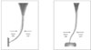
Sixty simulated resin root canal with a curvature of J and S-shape were divided into two groups. Each group consisted of three subgroups with 10 blocks according to the instruments used: ProFile®, ProTaper™, and K3TM. Simulated resin root canal was prepared by ProFile, ProTaper and K3 with 300 rpm by the crown-down preparation technique. Pre- and post-instrumentation apical foramen images were overlapped and recorded with Image-analyzing microscope 100X (Camcope, Sometech Inc, Korea). The amounts of difference in width and dimension on overlapped images were measured after reference points were determined by Image Analysis program (Image-Pro® Express, Media Cybernetic, USA). Data were analyzed using Kruskal-Wallis and Mann-Whitney U-test.
The results suggest that ProFile showed significantly less canal transportation and maintained original apical foramen shape better than K3 and ProTaper.
Figures and Tables
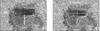 | Figure 2Procedure measuring the difference in width and dimensions of apical foramen. A. Measurement of difference in width of apical foramen after overlapping pre- and post-instrumentation images. (L1; Δ mesial width, L2; Δ outer width, L3; Δ distal width, L4; Δ inner width) B. Measurement of difference in dimensions of apical foramen after overlapping pre- and post-instrumentation images. (a; inner dimensions of apical foramen before instrumentation, b; outer dimensions of apical foramen before instrumentation, c; inner dimensions of apical foramen after instrumentation, d; outer dimensions of apical foramen after instrumentation)
|
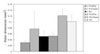
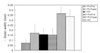 | Figure 3ΔOuter width of apical foramen between pre- and post-instrumentation according to the types of files and curvatures.
|
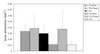
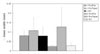 | Figure 4ΔInner width of apical foramen between pre- and post-instrumentation according to the types of files and curvatures.
|
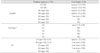
 | Figure 5ΔOuter dimensions of apical foramen between pre- and post-instrumentation according to the types of files and curvatures.
|
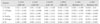
 | Figure 6ΔInner dimensions of apical foramen between pre- and post-instrumentation according to the types of files and curvatures.
|
References
1. Schilder H. Filling root canals in three dimensionss. Dent Clin North Am. 1967. 723–744.
2. Zmener O, Balbachan L. Effectiveness of nickel-titanium files for preparing curved root canals. Endod Dent Traumatol. 1995. 11:121–123.

3. Weine FS. Endodontic therapy. 1989. 4th ed. St. Louis: Mosby;277.
4. Walia H, Brantley WA, Gerstein H. An initial investigation of the bending and torsional properties of Nitinol root canal files. J Endod. 1988. 14:346–351.
5. Baumann MA. Nickel-titanium: options and challenges. Dent Clin North Am. 2004. 48:55–67.
6. Esposito PT, Cunningham CJ. A comparison of canal preparation with nickel-titanium and stainless steel instruments. J Endod. 1995. 21:173–176.
7. Park HS, Lee MG, Kim JJ, Lee JY. In the formation of curved root canals using ProFile Study on root canal shape. J Korean Acad Conserv Dent. 1999. 24:633–637.
8. Ko HJ, Baek SH. A study of histomorphological change of curved root canal preparation using GT rotary file, profile and stainless steel K-file. J Korean Acad Conserv Dent. 2002. 27:612–621.
9. Koch K, Brave D. The Ultimate Rotary File? Oral Health. 2002. 59–64.
10. Weine FS, Kelly RF, Lio PJ. The effect of preparation procedures on original canal shape and on apical foramen shape. J Endod. 1975. 1:255–262.
11. Hsu YY, Kim SK. The ProFile system. Dent Clin North Am. 2004. 48:69–85.
12. Clauder T, Baumann MA. ProTaper NT system. Dent Clin North Am. 2004. 48:87–111.
13. Mounce RE. The K3 rotary nickel-titanium file system. Dent Clin North Am. 2004. 48:137–157.
14. Bishop K, Dummer PMH. A comparison of stainless steel flexofiles and Nickel-titanium Ni-Ti flex during the shaping of simulated canals. Int Endod J. 1997. 30:25–34.
15. Coleman CL, Svec TA. Analysis of Ni-Ti versus stainless steel instrumentation in resin simulated canals. J Endod. 1997. 23:232–235.
16. Song YL, Bian Z, Fan B, Fan MW, Gutmann JL, Peng B. A comparison of instrument-centering ability within the root canal for three contemporary instrumentation techniques. Int Endod J. 2004. 37:265–271.
17. Short JA, Morgan LA, Baumgartner JC. A comparison of canal centering ability of four instrumentation tecniques. J Endod. 1997. 23:503–507.
18. Hata GI, Uemura M, Kato AS, Imura N, Novo NF, Toda T. A comparison of shaping ability using ProFile, GT file, and Flex-R endodontic instruments in simulated canals. J Endod. 2002. 28:316–321.
19. Lim JJ, Kim DJ, Hwang YC, Hwang IN, Oh WM. The change of canal configuration after instrumentation by several nickel-titanium files in the simulated canal with abrupt curvature. J Korean Acad Conserv Dent. 2005. 30:303–311.
20. Yun HY, Kim SK. A comparison of the shaping abilities of 4 nickel-titanium rotary instruments in simulated root canals. Oral Surg Oral Med Oral Pathol Oral Radiol Endod. 2003. 95:228–233.
21. Walsch H. The hybride concept of nickel-titanium rotary instrumentation. Dent Clin North Am. 2004. 48:183–202.
22. Peters OA, Peters CI, Schönenberger K, Barbakow F. ProTaper rotary root canal preparation: effects of canal anatomy on final shape analysed by micro CT. Int Endod J. 2003. 36:86–92.
23. Oh HJ, Hong CH, Jo YB. The effect of niti rotary instrumentation on the configuration of apical root canal. J Korean Acad Conserv Dent. 1997. 22:244–253.
24. Lee BK, Kim DJ, Hwang YC, Hwang IN, Oh WM. A comparative study on the canal configuration after shaping by profile, protapertm and k-flexofile in simulated canals with different angles of curvature. J Korean Acad Conserv Dent. 2005. 30:294–302.
25. Calberson FL, Deroose CA, Hommez GM, De Moor RJ. Shaping ability of ProTaper nickel-titanium files in simulated resin root canals. Int Endod J. 2004. 37:613–623.
26. Iqbal MK, Firic S, Tulcan J, Karabucak B, Kim S. Comparison of apical transportation between ProFile and ProTaper NiTi rotary instruments. Int Endod J. 2004. 37:359–364.
 XML Download