

 PDF
PDF ePub
ePub Citation
 Print
Print


I. Introduction
The loss of dental hard tissue at the cervical region of the tooth is a common clinical occurrence. These noncarious cervical lesions (NCCLs) present in a variety of forms1,2). Traditionally, the most commonly cited etiological factors thought to lead the development of NCCLs are erosion, abrasion.
An additional mechanism for cervical tooth structure loss with occlusal loading has been proposed. It is suggested that occlusal loads applied during lateral excursion cause the tooth to flex. As the tooth flexes, these stresses may cause disruption of the bonds between the hydroxyapatite crystals leading to enamel loss. This type of hard tissue loss at the cemento-enamel junction has been termed 'abfraction' by Grippo to distinguish it from lesions caused by erosion and abrasion3). The abfraction is accelerated by excessive occlusal load such as occlusal interferences, premature contacts, bruxism, and clenching habit. Clinically, abfraction lesions are sharp, angular, wedge shaped defects that sometimes have a subgingival location and are often associated with marked wear facets4,5).
When a patient is experiencing pain or hypersensitivity and food impaction in lesion or concerned about the unesthetic appearance, it is the indication of restoration and potential for tooth fracture or pulp exposure are also reasons for restoration6). Grippo7) recommended restoration in order to prevent further enlargement of the lesion.
There are a number of materials that can be used to esthetically restore abfraction lesions. Tooth-colored direct restorative materials such as glass ionomer cement, composite resins, and resin-modified glass ionomer are available8).
The most appropriate one is composite resin, in spite of some disadvantages like time-consuming, tech-sensitivity, volumetric shrinkage, which can also exhibit superior esthetics, polishability, wear resistance. Blunck8) recommended the composite resin for wedge shaped lesions because of the long-term clinical appearance seems to be superior to other materials. Some authors9-11) suggested that the use of more flexible restorative materials like compomers and filled bonding agents may be able to resist shear forces generated by tooth flexure and therefore enhance retention. And Ausiello et al.12) reported that the use of a thin layer of a more flexible adhesive (with lower elastic modulus) exhibits the same rigidity as thick layers of less flexible adhesive (with higher elastic modulus). The complex biomechanical behavior might arise from the simultaneous effects of polymerization shrinkage, composite stiffness and adhesive interface strain12). And the elastic modulus of the used material and the adhesive layer properties may be important factors in restoration durability. Clinically, in order to solve these problems, sandwich technique may be introduced in the restoration of deep NCCL with the glass ionomer lining which has lower elastic modulus and no shrinkage13).
Finite element analysis (FEA) is a mathematical method in which the geometry of a specimen is divided into a sequence of distinct elements interconnected at nodes. It is particularly useful in dentistry since it can readily cope with both the complex geometry of a tooth and it's supporting structures and with the large variation in the physical properties of the tooth, periodontal ligament and alveolar bone14).
A few investigations15-17) about NCCLs were made using FEA and these researches generally were conducted using two-dimensional (2D) analysis for vertical section of class V cavity. There were several studies of 3D FEA about class V cavity with regard to polymerization shrinkage by light curing18,19). But most studies about the stress distribution of class V cavity didn't consider multi-factors, such as restorative material, polymerization shrinkage and occlusal loading condition, simultaneously.
The purpose of this study was to investigate the influence of elastic modulus, polymerization shrinkage of restorative materials and occlusal loading condition on the stress distribution of notch shaped NCCL using 3D FEA.
II. Materials and methods
FE model
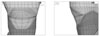
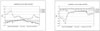
To developing a 3D FE model, an intact normal extracted human maxillary second premolar was used in this study. The extracted premolar was scanned serially with Micro-CT (SkyScan1072; SkyScan, Aartselaar, Belgium) to expose the tooth sections perpendicular to the long axis of the tooth (58 µm in thickness) and parallel to the occlusal plane. Image processing software, 3D-DOCTOR (Able Software Co., Lexington, MA, USA), was employed to make the boundaries of enamel, dentin and pulp and construct a surface model of tooth from the sectioned two dimensional images. Rhino3D (Robert McNeel & Assoc., Seattle, WA, USA) was used to reduce useless nodes from the surface model and ANSYS (Swanson Analysis Systems, Inc., Houston, USA) was used to mesh.
The final model consisted of 16668 elements with 18245 nodes and the restored model had 17636 elements and 19012 nodes.
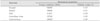
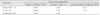
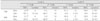
All the vital tissues were presumed linearly elastic, homogeneous and isotropic. The corresponding elastic properties such as Young's modulus and Poisson's ratio were determined according to literature survey (Table 1). The periodontal ligament was assumed to be 0.3 mm wide, and the dimensions of surrounding compact and cancellous bone were derived from standard texts. The alveolar bone was also generated by growing the outer surface of the tooth model from 2 mm below the cemento-enamel junction (CEJ)20,21). The pulp region was modeled as being hollow22). In these models, the outer surface of the alveolar bone model was fixed in order to prevent rigid body motion for FEA.
The model was fixed on mesiodistal direction. In all loading cases, the base nodes of simulated alveolar bone were assumed fixed to prevent rigid body motion.
Experimental conditions and simulated groups
Loading conditions
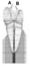
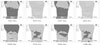
A static load of 500 N was applied on a point load condition at buccal and palatal cusps. Perpendicular load on the upper third of the palatal slope of the buccal cusp (Load A) and perpendicular load on the upper third of the buccal slope of the palatal cusp (Load B) were used in this study (Figure 1).
Restorative materials and adhesive layer thickness
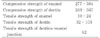
Notch shaped cavity was filled with either hybrid or flowable resin and each restoration was simulated with a adhesive layer thickness (40 µm) (Figure 3). The data of material properties such as elastic modulus, Poisson's ratio and polymerization shrinkage were determined by literature review (Table 2). The Tetric Flow (Vivadent Ets., FL-9494-Schaan, Liechtenstein) and Z100 (3M Dental Products, St. Paul, MN, USA) were used as representatives of flowable and hybrid resin. Dentin bonding system used in this study was Scotchbond Multi Purpose (3M Dental Products, St. Paul, MN, USA). The adhesive layer was made by mathematical shell element modeling and the conjunctions between materials were set as complete coupling (Figure 2).
Groups for simulation
The variables were loading condition (A or B) and restoration material (Tetric Flow; T or Z100; Z). The tested groups were classified as 4 situations by the restoration material (T or Z) and loading condition (A or B) with adhesive layer thickness (40 µm); T40A, T40B, Z40A, Z40B.
Stress analysis
After shear stress set to be zero, normal stress was expressed as S1, S2 and S3. And then, S1 was appointed as maximum principal stress and S3 as minimum principal stress. A graph was obtained based on the data of principal stresses at each node with load A or B.
The principal stresses in the lesion apex (that is internal line angle of cavity) and middle vertical wall were analyzed using ANSYS.
III. Results
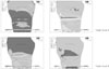
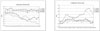
Principal stresses before restoration (Figures 3, 4 and Table 4)
Before restoration, the stresses were highly concentrated at mesial CEJ and lesion apex (Figure 3).
The first peak stresses of tensile stress at all area of notch shaped NCCL were over the mechanical properties of teeth.
The peak compressive stress of 588.0 MPa was concentrated at mesial point angle (MP) of the lesion under load A and the peak tensile stress of 193.3 MPa was concentrated at MP under load B.
Principal stresses after restoration
After restoration using Z100 and Tetric Flow, there was significant stress relief, but its magnitude was different for each area and each treatment (Figure 5).
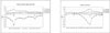
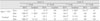
Lesion apex and CEJ (Figure 6 and Table 5)
On lesion apex and CEJ, the peak compressive stress of 299.1 MPa was concentrated at M1 point under load A by restoration using T40. By restoration using Z100, the relatively low compressive stress of 233 MPa was concentrated at M1 point.
The peak tensile stress of 93.2 MPa was concentrated at M1 point under load B by restoration using T40. By restoration using Z100, the relatively low tensile stress of 68 MPa was concentrated at M1 point.
Middle vertical cavity wall (Figure 7, Table 6)
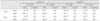
On middle cavity wall, the peak compressive stress of 70.8 MPa was concentrated at cervical cavosurface margin under load A by restoration using Z40. By restoration using T40, the relatively low compressive stress of 57.4 MPa was concentrated at lesion apex.
The peak tensile stress of 39.9 MPa was concentrated at the node of 17 under load B by restoration using Z40. By restoration using T40, the relatively low tensile stress of 31.7 MPa was concentrated at the node of 15.
IV. Discussion
The notch shaped lesion has a sharp line angle at the apex of the lesion, and the location with the highest stress concentration corresponded to sharpest geometric discontinuity. Therefore, it is suggested that the severity of geometric discontinuity of NCCL plays a significant role in the development of internal stress in tooth1-5). Since cervical lesions act as stress concentrators, NCCL, especially abfraction lesions, will extend if not restored. Grippo also suggested that, if the lesion is left unrestored, the stress concentration caused by the cervical lesion would facilitate further deterioration of tooth structure. He also hypothesized that restoration decrease the concentration of stress and further progress of the lesion7). But Osbome-Smith et al.27) concluded that the restoration of NCCL with resin-modified glass ionomer or composite resin did not result in a statistically significant increase in the fracture resistance.
Kuroe et al.28) reported that, regardless of lesion shape, higher stress was developed at the apex of an unrestored cervical lesion than at any other portion of the tooth when the cusp located above the lesion was loaded. In this study, the stress distributions were analyzed with 3D FEA with the variables of loading condition, the thickness of adhesive layer and restorative material. According to stress analysis, mesial CEJ corner and lesion apex showed high stress distribution before restoration. It is expected that unrestored notch shaped NCCL would be extended around the mesial and distal corner area of all cavosurface margin by continuous failure with excessive compressive or tensile stress under load A or load B, respectively. Because of the anatomical mesiodistal asymmetry, higher stresses were concentrated at mesial point angle than distal.
Other than wear resistance and color stability, the most important characteristic of restorative material is the modulus of elasticity or stiffness of the material. This is important for the masticatory stresses are transferred through the cusp and concentrated at the cervical region. It is recommended, therefore, that composite resin with a low modulus of elasticity be used. With this type of resin, much of the transferred energy is absorbed by the restoration rather than transmitted to the dentin-restoration interface29).
In a study of various resin-based restoratives, Yaman et al.30) concluded that if the restoration material had larger Young's modulus, less stress was developed in restored tooth. Similar to the results of this study, Z100 was found to be superior to the other materials concerned. And they revealed that by increasing load size or load angle or cavity size, there was increase in stress distribution30). On the other hand, there was an investigation that no differences were found between a microfilled and a hybrid resin, although two materials have widely different moduli of elasticity31). As the results of this study, the principal stresses were remarkably reduced at highly stress concentrated area such as lesion apex or CEJ with restoration using Z100, while they were reduced on the other areas with restoration using Tetric Flow. This was the same results by Yaman30) that restoration using Z100 worked as a strut to prevent stress concentration of the lesion.
After restoration, highly stress concentrated area migrated from mesial point angle to mesial CEJ near the point angle. This might be the reason of progression of the lesion to CEJ area, especially in Tetric Flow restoration.
As the results of this study, compressive stresses were created under load A and tensile stresses under load B in the point of principal stresses. The peak stresses of both principal stresses were decreased after restoration, but high stress concentration still remained around mesial point angle. This compressive stresses over the failure range under load A could be decreased under the limit after restoration, but tensile stresses over the limit range under load B were still remained on various areas. These mean that the tensile stresses may be the major factor to jeopardize the restoration durability and to promote the lesion progression. Therefore some clinicians28,32,33) recommended that occlusal adjustment be considered as a treatment option, in addition to restoration, for abfraction lesions. Occlusal adjustments may reduce stress concentration at the cervical lesion, but they may jeopardize equilibrium of the occlusion if careful consideration for the entire occlusion is omitted. And they are also difficult to do with confirm on which side of occlusion concerning tensile or compressive is modified. Therefore the restoration of NCCL is first concern and then the selection of restorative material may have influence on the prognosis of the lesion and restoration.
In future, it is recommended to evaluate the restoration of NCCL using not only a single material but also two or more materials, based on the relation of restorative material's mechanical properties and regional differences of stresses in the lesion. And the selection of the mechanical properties of the restorative material for long-term clinical success is the question remaining to be answered.
V. Conclusion
1. Under loading A, compressive stress is created in the unrestored and restored cavity. Under loading B, tensile stress is created. And the peak stress concentration is seen at near mesial corner of the cavity under each load condition.
2. Compared to the unrestored cavity, the principal stresses at the CEJ and line angle of the cavity were more reduced in the restored cavity on both load condition.
3. In teeth restored with hybrid composite, the principal stresses at the CEJ and line angle of the cavity were more reduced than flowable resin.
 XML Download