

 PDF
PDF ePub
ePub Citation
 Print
Print


I. INTRODUCTION
Three dimensional root canal shaping and its hermetic obturation are the major elements determining the predictability of successful endodontics.
Root canal instruments manufactured with nickel-titanium (NiTi) alloys have been developed in an attempt to overcome the limitations of them made with stainless steel alloys1). NiTi rotary instruments have helped the root canal to be prepared easier and faster than hand instruments2). And the use of NiTi rotary instrumentation can effectively produce a well-tapered root canal form sufficient for obturation with minimal risk of transporting the original canal, and are helpful in the preparation of curved root canals2-6).
It is estimated that there are more than least 18 different rotary file brands in the endodontic marketplace7). Most of these NiTi file systems - e.g. ProFile (Dentsply Maillefer, Ballaigues, Switzerland), K3 (SybronEndo, Glendora, France), Mtwo (VDW, Munich, Germany), Hero642 (Micromega, Besancon, France) and Hero Shaper (Micromega, Besancon, France), - have a constant tapered shaft design, while these have their own rake angles and radial lands6,8-10).
Distinctively, the ProTaper system (Dentsply Maillefer, Ballaigues, Switzerland) is an unique instrument which has progressively changing tapered shaft design along a single instrument in the shaft design11,12). And the design of cross-section of ProTaper - convex triangular - is claimed to reduce the contact area between the file and dentin, and to have excellent cutting efficiency12). It was claimed that the ProTaper system provides a continuous tapered preparation of the root canal, without significant transportation of the original curvature of the root canal12). And also it was suggested that the ProTaper instruments might be better suited for curved and constricted canals than for wide, immature ones12,13).
However, since the ProTaper system has been introduced, the possibility of transportation in the root canal prepared by active cutting action was discussed in more or less severe curved canals. Several studies showed that ProTaper instruments produced more aberrations, deformation and straightening of the canal14-16). Peter et al.13) showed that the ProTaper system tends to transport canals slightly larger than other file systems with a passive cutting action by evaluating shaped canal studies with micro CT. Lee et al.17) demonstrated that the ProTaper files remove too much canal structure and cause severe canal transportation than other files by the study using resin blocks.
And then, there were some reports suggesting that the NiTi files of less taper or U-file designs (e.g. ProFile) are good for refining of the apical portion of small curved canals because of its greater elasticity18-20).
Therefore, the combined use of the ProTaper with other file systems can emphasize the advantages of each system. This hybrid concept combining different file systems and different instrumentation techniques suitable for individual clinical situations, enables the clinicians to achieve the best biomechanical cleaning and shaping results more easily and simply19,20).
Recently, new ProTaper system (ProTaper Universal) with different configuration and cross-sectional design have been marketed. The ProTaper Universal has the same triangular cross-sectional design in S series and F1, but has modified cross-sectional design in F2 and F3 in which had triangular cross-section of notched side. And these instruments are characterized by the lower conicity in the upper part of the file to enhance the tactile feeling while preparing the apical portion of the canals. In addition, the manufacturer insisted that ProTaper Universal instrument has more flexible character inherently along the whole length of the files.
The purpose of this study is to compare and evaluate the shaping abilities of ProTaper, ProTaper Universal system, and two hybrid instrumentation methods using S series of ProTaper Universal and Hero Shaper or ProFile.
II. MATERIALS AND METHODS
1. The simulated root canals and pre-instrumented image taking
One hundred simulated root canals in clear resin blocks (Endo Training Bloc; Dentsply Maillefer, Ballaigues, Switzerland) were used for this study. The mean length of the root canals were 17 mm and the mean curvature of the canals were 40°.
Aqueous methylene blue dye was injected into the canal to enhance the contrast of the pre-instrumentation image. These resin blocks were scanned in a reproducible position with a scanner (Scanjet C8510A; Hewlett-Packard, California, USA), and then the data were stored in a personal computer.
2. Classification of groups and preparation
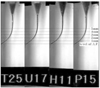
The simulated canals were divided into four groups according to the used instrument and method (Table 1). Each group had 25 resin blocks.
The experienced clinician in endodontic treatment who did it for about 10 years prepared the simulated root canals. All of the rotary NiTi files were operated by an electric motor (X-smart; Dentsply Maillefer, Ballaigues, Switzerland) set at 300 rpm and torque of 2.4 N. These setting values were within the range suggested at the manufacturer's operation manuals. The use of files was also in accordance with the manufacturer's recommendation. All files used in this study were new ones.
During the preparation procedures, canals were verified the patency with #10 K-Flexofile (Dentsply Maillefer, Ballaigues, Switzerland) then irrigated with saline and lubricated with RC-prep (Stone Pharmaceuticals, Philadelphia, USA) repeatedly. The root canals were copiously irrigated with saline dispensed through a 27-gauge needle (ENDO-EZE; Ultradent, South Jordan, USA) after using each instrument. After filing, apical gauging was verified with a precurved #30 K-Flexofile.
The instrumentation procedures were summarized at Table 2.
Group T
ProTaper instruments of S1, S2, F1, F2, and F3 were used in crown-down manner according to the manufacturer's instructions using a gentle in-and-out motion.
Group U
ProTaper Universal instruments of S1, S2, F1, F2, and F3 were used in crown-down manner according to the manufacturer's instructions using a gentle in-and-out motion.
3. Post-instrumented image taking and superimposition
After the root canal preparation was completed, the enlarged canal was filled with Vitapex (Neo Dental Chemical Products co., LTD, Tokyo, Japan) to enhance the contrast of the post-instrumentation image. Then the resin block was scanned again at the reproducible position. The pre- and post-instrumented canal images were superimposed on one another with the guide of attached label using Adobe Photoshop ver.7.0 (Adobe, San Jose, California, USA) (Figure 1). These superimposed images of 2736 × 944 pixel size were inspected on the TFT-LCD monitor (SyncMaster CX1565N; Samsung, Suwon, Korea) at 1024 × 768 screen resolutions.
4. Measurement and calibration
1) Increased canal width
The width of the canal after instrumentation was measured linearly from the pre-instrumented point to the post-instrumented wall inwardly and outwardly. Measurements were taken horizontally at 1, 2, 3, 4 and 5 mm from the level of apical foramen. Internal and external instrumented widths were summed to get the increased canal width.
2) Apical centering ratio
The centering ratio was to evaluate the movement of root canal center after preparation; the smaller the ratio, the better the instrument remained centered in the canal11,21,22). The ratio was computed using the following method: the absolute value of a net transportation (difference between internal and external instrumented width) divided by the whole width of the post-instrumented canal.
3) Instrumentation time
Instrumentation time was recorded in seconds by one assistant. The time for irrigation, changing instruments and patency was excluded to measure only shaping time.
4) Canal aberrations
Assessments were made according to the presence of various types of canal aberrations such as apical zip, elbow and ledge using the scanned post-instrumented images.
5) Instrument distortion
After root canal preparation, the NiTi files were verified for permanent distortion under the operative microscope (OPMI pico; Carl Zeiss, Oberkochen, Germany) at 25-fold magnification.
6) Reduction of root canal curvature and change of radius of root canal curvature
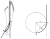
The degree of the root canal curvature may be diminished after instrumentation by the root canal straightening. The root canal curvatures of before and after root canal preparation were measured by Schneider method23). And the decreased value of root canal curvature after root canal preparation was calculated (Figure 2).
Generally, the radius of curvature is lengthened after instrumentation due to the root canal straightening. The radius of curvature was calculated on the basis of the geometric principals of an isosceles triangle23). It was possible to calculate the radius according to the following formula: r = s / 2 sin A (Figure 2).
5. Statistical analysis
Statistical analysis of the collected data was performed with SPSS version 10.0 (SPSS Inc., Chicago, IL, USA). Mean scores of each group were statistically analyzed using ANOVA and Duncan's multiple range test for post-hoc comparison. The Pearson Chi-Square test was done to compare the frequencies of canal aberration and instrument distortion. Differences revealed in the data were designated as significant at P < 0.05.
III. RESULTS
1. Increased canal width
The representative superimposed images of each group are presented in Figure 3.
Generally, the increased canal width in Group U was wider than in Group H and Group P, but narrower than in Group T except 1 mm level where the two ProTaper groups cut similar amount of canal wall (Table 3). The width with ProFile was narrower than other instruments in all level. The increased canal width of ProTaper and ProTaper Universal group were larger than that of Hybrid instrumentation groups (P < 0.05).
2. Apical centering ratio
The centering ratio of each level showed significant difference between groups (Table 4, P < 0.05).
At 1 mm level, Group P was d istinctive from other groups. Group P had the smallest centering ratio of all the groups (P < 0.05). At 2 mm level, Group H had significantly larger centering ratios than other groups (P < 0.05), and at 3 mm level there was no significant differences between groups. At 4 mm level, the two hybrid methods showed better centering ratio (P < 0.05). At 5 mm level, although there was no significant difference between Group T and Group H, the two hybrid methods showed better centering ratio than Group T and Group U.
3. Instrumentation time
The time needed for completion for each system is presented in Table 5. The hybrid instrumentation using ProFile needed more time than other system.
4. Instrument distortion
There were only three files distorted. Two .04 #30 Hero Shaper and a .04 #25 ProFile showed permanent deformation. There was no broken file after instrumentation.
6. Reduction of root canal curvature and increase of radius of root canal curvature
There was no significant difference in the reduction of root canal curvature and the increase of radius of root curvature between groups (Table 6, P > 0.05).
IV. DISCUSSION
During the last few years the number of users has been increased significantly and many new systems were launched on the market.
Each file system has its own special advantages and disadvantages, and particular rules for its operation. Likewise, ProTaper has many reports regarding its usefulness in the shaping of coronal and mid-root portion12,18,19). Despite its several advantages, however, a few studies showed that the ProTaper instruments produced more aberration, deformation and straightening of the canal especially at the apical portion, due to their thicker and stiffer finishing files of F1, F2 and F314-16). Because of these possibilities of the procedural errors, Berutti et al.18) and Walsch19) recommended that the use of passive or manual NiTi files, because they might be better for the final phase of shaping curved canals as a hybrid technique.
The constant tapered shaft design of the NiTi file is more flexible than the progressive tapered shaft design. And the U-shaped cross section is more flexible than the triangular cross section11,12). Based on the comparative report of mathematical models of ProTaper and ProFile, the results have shown that the ProTaper might be more indicated for narrow canals and curved canals during the initial phase of shaping and that the ProFile might be more indicated in the final phase of shaping18). Hong et al.24) recommended that the hybrid technique using the ProTaper S1 combined with ProFile or Hero series. Especially in novice operator, the combined usage of ProTaper and ProFile was shown to have the better apical centering ratio than ProTaper alone and other hybrid method using stainless steel file or Hero Shaper.
The manufacturer insisted that potential short-comings by the ProTaper such as apical transportation could be minimized by the geometric modification in ProTaper Universal. This study was performed with another intention to verify that the ProTaper Universal might have actual improvement of lesser transportation than ProTaper, and that consequently the ProTaper Universal decreases the necessity of hybrid technique.
Thus, the conventional ProTaper and ProTaper Universal systems were compared with the two hybrid instrumentation methods combining S series of ProTaper Universal and other instruments. The S1 and S2 files of ProTaper Universal were used for the performance of body shaping and apical pre-enlargement, and the respective subsequent system of Hero Shaper and ProFile was operated for the shaping and refining of the apical portion.
The instrumented width is determined by the instrument size inherently and influenced by the amount of deviation. Although several researches13,15,16) about ProTaper reported the large centering ratio and transportation tendency after completion of root canal preparation at apical portion, the ProTaper Universal showed lesser increased width than ProTaper in this study. It is assumed that the ProTaper Universal would make lesser deviation or transportation. But it still showed that the more deviation or transportation might happen than ProFile and Hero Shaper. The differences observed between Groups P and H may be explained by the geometric design of ProFile and Hero Shaper. With regards to the instrument design, Powell et al.25) has reported that the tip design of the instrument affects the shape of canals and the levels of deviation. Generally, active instruments cut more effectively and more aggressively with the tendency to straighten the canal curvature26). In curved canals, canal transportation might be formed because of not only instrument design but also other factors, such as the canal curvature, physical properties of alloy, and techniques22,27).
The centering ratio was used as an index of the capability of the instrument to stay centered in the canal; the smaller the ratio, the better the instrument remained centered in the canal21,22).
The ProTaper Universal showed similar centering ratio with ProTaper in all levels and there was no improvement. Interestingly, at 1 mm and 3 mm levels, the two ProTaper systems were not significantly different from Hero Shaper system. Particularly, at 2 mm level the two ProTaper systems showed significantly lesser centering ratios than Group H. The ProFile had the better centering ability than the other groups in most of evaluated levels. This finding is consistent with other studies to evaluate the centering ratio or shaping ability of various NiTi files including ProFile24,28). Hong et al.24) recommended, through the comparison of various hybrid instrumentation technique, that if the apical regions are to be enlarged wider than ISO 30, it might be better to use a different system12). But this study showed that all experimental system makes similar centering ratio except Group P. Schäfer et al.16) reported that ProTaper tended to transport towards the outer aspect of the canal curvature. Peters et al.13) reported that ProTaper design may also increase the incidence of procedural errors and overall canal transportation whilst the modified cutting flute of ProTaper instruments may reduce friction and consequently reduce the torque. But no group had made the canal aberrations in this test. Properly used NiTi file systems enable the user to finish more predictable root canal instrumentation and limit procedural errors at the same time12).
A relevant aspect of the described radii is that the susceptibility to fracture of rotary nickel-titanium instruments is affected by the radius of the curved canal29,30). The cyclic fatigue of rotary nickel-titanium instruments has been investigated by using simulated canals with radii of 10 mm and 5 mm30) and 5 mm and 2 mm29); in both studies it was noted that as the radius of curvature decreased, cycles to failure decreased. Radius of curvature was decided to be the most significant factor for determining the fatigue resistance of these instruments30).
Generally, the root canal curvature will be diminished and the radius of curvature will be increased after preparation. In this study, there were no significant differences in curvature and radius change between the shaping methods.
Clinicians do not want to spend lots of time for root canal preparation. It usually took shorter time to prepare the canals with the ProTaper systems than other NiTi rotary file systems in other studies15,16). The instrumentation time with Group U was shorter than Group T and similar with Group H. It seemed that the changed design of ProTaper Universal improved the time lapse for shaping. Although the hybrid methods in this study needed one more file than ProTaper systems, Group H showed similar preparation time. The lesser differences between groups might come from the operator's proficiency. And the frequency of file binding may affect the instrumentation time and it might be also influenced by the operator's proficiency.
One of the main problems inherent in the rotary preparation by clinicians is the risk of instrument fracture27). In this study, there was no fracture of instruments. However the distortions that might lead to fracture and hinder ideal canal preparation were noted in Group P and Group H. Despite the notice, no statistical conclusions could be made by reason of the low frequency of occurrence. The operator's proficiency, undoubtedly, is a significant factor in reducing fracture incidence of NiTi rotary instruments and root canal aberrations.
Most breakage of rotary files, regardless of the manufacturer, is because the dentist failed to follow the directions for that particular file. It has been said that every dentist has the possibility of breaking every rotary file. The most important thing, therefore, is not the kinds of the file; it is, as always, the clinician7). To increase safety, most endodontic teachers and educators emphasize sufficient supervised training by endodontists that have mastered any particular system7).
The hybrid methods of each the ProFile and Hero Shaper system with ProTaper S1 are recommendable comparative to ProTaper alone method in the study of Hong et al.24), however, according to the present results, we can suggest the ProTaper Universal alone may be an appropriate method comparable with hybrid techniques in curved root canal preparation.
V. CONCLUSION
Under the conditions of this study, the ProTaper Universal seems to have better shaping ability than ProTaper in terms of instrumented width and instrumentation time. It can be suggested that hybrid instrumentation using ProTaper and other constant-tapered NiTi file is not efficient anymore in highly experienced operators. The experts can do appropriate root canal shaping with not only hybrid technique but also ProTaper Universal system.


 XML Download