

 PDF
PDF ePub
ePub Citation
 Print
Print


Abstract
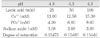
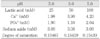
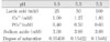
The purpose of this study is to compare and to evaluate the effect of pH and lactic acid concentration on the progression of artificial root caries lesion using polarizing microscope, and to evaluate the morphological changes of hydroxyapatite crystals of the demineralized area and to investigate the process of demineralization using scanning electron microscope.
Artificial root caries lesion was created by dividing specimens into 3 pH groups (pH 4.3, 5.0, 5.5), and each pH group was divided into 3 lactic acid concentration groups (25 mM, 50 mM, 100 mM). Each group was immersed in acid buffer solution for 5 days and examined. The results were as follows:
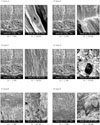
1. Under polarized microscope, the depth of lesion was more effected by the lactic acid concentration rather than the pH.
2. Under scanning electron microscope, dissolution of hydroxyapatite crystals were increased as the lactic acid concentration increased and the pH decreased.
3. Demineralized hydroxyapatite crystals showed peripheral dissolution and decreased size and number within cluster of hydroxyapatite crystals and widening of intercluster and intercrystal spaces as the pH decreased and the lactic acid concentration increased.
4. Under scanning electron microscope evaluation of the surface zone, clusters of hydroxyapatite crystals were dissolved, and dissolution and reattachment of crystals on the surface of collagen fibrils were observed as the lactic acid concentration increased.
5. Under scanning electron microscope, demineralization of dentin occurred not only independently but also with remineralization simultaneously.
In conclusion, the study showed that pH and lactic acid concentration influenced the rate of progression of the lesion in artificial root caries. Demineralization process was progressed from the surface of the cluster of hydroxyapatite crystals and the morphology of hydroxyapatite crystals changed from round or elliptical shape into irregular shape as time elapsed.
Figures and Tables
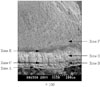
Figure 1
Polarizing microscopic view of demineralized dentin of group pH 4.3 in acid buffer solution (5 day treatment, × 100, imbibed in water).
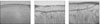
Figure 2
Polarizing microscopic view of demineralized dentin of group pH 5.0 in acid buffer solution (5 day treatment, × 100, imbibed in water).
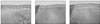
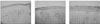
Figure 3
Polarizing microscopic view of demineralized dentin of group pH 5.5 in acid buffer solution (5 day treatment, × 100, imbibed in water).
References
1. Darby ET. The etiology of caries at the gum margin and labial and buccal surfaces of the teeth. Dental Cosmos. 1984. 26:218–232.
2. Katz S, Park KK, Palenik CJ. In vitro root surface caries studies. J Oral Med. 1987. 42:40–48.
3. Katz RV, Hazen SP, Chilton NW, Mumma RD Jr. Prevalence and intraoral distribution of root caries in an adult population. Caries Res. 1982. 16:265–271.

4. Beck JD, Hunt R, Hands JS, Field HM. Prevalence of root and coronal caries in a noninstitutionalized older population. J Am Dent Assoc. 1985. 111:964–968.
5. Tohda H, Fejerskov O, Yanagisawa T. Transmission electron microscopy of cementum crystals correlated with Ca and F distribution in normal and carious human root surfaces. J Dent Res. 1996. 75:949–954.
6. Hayashi Y. High resolution electron microscopy of enamel crystallites demineralized by initial dental caries. Scanning Microsc. 1995. 9:199–205.
7. Holmen L, Thylstrup A, Featherstone JDB, Fredebo L, Shariati M. A scanning electron microscopic study of surface changes during development of artificial caries. Caries Res. 1985. 19:11–21.
8. Carter WJ, Dunn JK, Fosdick LS, Moore BW. The formation of lactic acid in dental plaque. J Dent Res. 1956. 35:778–785.
9. Theuns HM, van Dijk JWE, Driessens FCM, Groeneveld A. Effect of the pH of buffer solutions on artificial carious lesion formation in human tooth enamel. Caries Res. 1984. 18:7–11.
10. Theuns HM, van Dijk JWE, Driessens FCM, Groeneveld A. Effect of time, degree of saturation, pH and acid concentration of buffer solutions on the rate of in vitro demineralization of human enamel. Arch Oral Biol. 1985. 30:37–42.
11. Margolis HC, Zhang YP, Lee CY, Kent RL Jr, Moreno EC. Kinetics of enamel demineralization in vitro. J Dent Res. 1999. 78:1326–1335.
12. Margolis HC, Murphy BJ, Moreno EC. Development of caries-like lesions in partially saturated lactate buffers. Caries Res. 1985. 19:36–45.
13. Silverstone LM, Johnson NW, Hardie JM, Williams RA. Dental caries, aetiology, pathology and prevention. 1981. the MacMillan Press;176–177.
14. Aamdal-Scheie A, Luan WM, Dahlen G, Fejerskov O. Plaque pH and microflora of dental plaque on sound and carious tooth surface. J Dent Res. 1996. 75:1901–1908.
15. Feagin FF, Graves CN. Evaluation of the effects of F in acidified gelatin gel on root surface lesion development in vitro. Caries Res. 1988. 22:145–149.
16. Hoppenbrouwers PM, Driessens FCM, Borggreven JMPM. The vulnerability of unexposed human dentin roots to demineralization. J Dent Res. 1986. 65:955–958.
17. Hoppenbrouwers PM, Driessens FCM, Borggreven JMPM. The mineral solubility of human tooth roots. Arch Oral Biol. 1987. 32:319–322.
18. Shellis RP. Effects of a supersaturated pulpal fluid on the formation of caries-like lesions on the roots of human teeth. Caries Res. 1994. 28:14–20.
19. Featherstone JD. Diffusion phenomena during artificial carious lesion formation. J Dent Res. 1977. 56 Special D:D48–D52.
20. Soni NN, Brudevold F. Microradiographic and polarized-light studies of artificially produced lesions. J Dent Res. 1960. 39:233–240.
21. Clarkson BH, Hall DL, Heilman JR, Wefel JS. Effect of proteolytic enzymes on caries lesion formation in vitro. J Oral Pathol. 1986. 15:423–429.
22. Clarkson BH, Wefel JS, Miller I. A model for producing caries-like lesions in enamel and dentin using oral bacteria in vitro. J Dent Res. 1984. 63:1186–1189.
23. Clarkson BH, Krell D, Wefel JS, Crall J, Feagin FF. In vitro caries-like lesion produced by Streptococcus mutans and Actinomyces viscosus using sucrose and starch. J Dent Res. 1987. 66:795–798.
24. Shay K. Root caries in the older patient. Dent Clin North Am. 1997. 41:763–793.
25. Silverstone LM. Observations on the dark zone in early enamel caries and artificial caries-like lesions. Caries Res. 1967. 1:260–274.
26. Moreno EC, Zahradnik RT. Chemistry of enamel subsurface demineralization in vitro. J Dent Res. 1974. 53:226–235.
27. Wefel JS, Heilman JR, Jordan TH. Comparisons of in vitro root caries models. Caries Res. 1995. 29:204–209.
28. Phankosol P, Ettinger RL, Hicks MJ, Wefel JS. Histopathology of the initial lesion of the root surface: an in vitro study. J Dent Res. 1985. 64:804–809.
29. Schüpbach P, Guggenheim B, Lutz F. Human root caries - histopathology of initial lesions in cementum and dentin. J Oral Pathol Med. 1989. 18:146–156.
30. Wefel JS, Clarkson BH, Heilman JR. Natural root caries - a histologic and microradiographic evaluation. J Oral Pathol. 1985. 14:615–623.
31. Furseth R, Johansen E. The mineral phase of sound and carious human dental cementum studied by electron microscopy. Acta Odontol Scand. 1970. 28:305–322.
32. Westbrook JL, Miller AS, Chilton NW, Williams FL, Mumma RD Jr. Root surface caries - a clinical, histopathologic and microradiographic investigation. Caries Res. 1974. 8:249–255.
33. Takuma S, Ogiwara H, Suzuki H. Electron-probe and electron microscope studies of carious dentinal lesions with a remineralized surface layer. Caries Res. 1975. 9:278–285.
34. LeGros RZ. Formation and stability of synthetic apatites. Calcium phosphates in oral biology and medicine. 1991. Karger;82–107.
35. Buskes JA, Christoffersen J, Arends J. Lesion formation and lesion remineralization in enamel under constant composition conditions - A new technique with applications. Caries Res. 1985. 19:490–496.
36. Han WS, Lee CY. The effects of the fluoride concentration of acidulated buffer solutions on dentine remineralization. 2004. Yonsei University.
37. Schüpbach P, Guggenheim B, Lutz F. Histopathology of root surface caries. J Dent Res. 1990. 69:1195–1204.
38. Schüpbach P, Guggenheim B, Lutz F. Human root caries - histopathology of advanced lesions. Caries Res. 1990. 24:145–158.
39. Schüpbach P, Guggenheim B, Lutz F. Human root caries - histopathology of arrested lesions. Caries Res. 1992. 26:153–164.
40. Nyvad B, ten Cate JM, Fejerskov O. Microradiography of experimental root surface caries in man. Caries Res. 1989. 23:218–224.
41. Nyvad B, Fejerskov O. Root surface caries - clinical, histopathological and microbiological features and clinical implications. Int Dent J. 1982. 32:311–326.
42. Nyvad B, Fejerskov O. Thylstrup A, Leach SA, Qvist V, editors. Active and inactive surface caries - structural entities? Dentine and dentine reactions in the oral cavity. 1987. Oxford: IRL Press;165–179.
43. Daculsi G, Kerebel B, Le Cabellec M, Kerebel L. Qualitative and quantitative data on arrested caries in dentine. Caries Res. 1979. 13:190–202.
44. Daculsi G, Le Geros RZ, Jean A, Kerebel B. Possible physico-chemical processes in human dentin caries. J Dent Res. 1987. 66:1356–1359.
45. Arends J, Christoffersen J. The nature of early caries lesions in enamel. J Dent Res. 1986. 65:2–11.
46. Dietz W, Kraft U, Hoyer I, Klingberg G. Influence of cementum on the demineralization and remineralization processes of root surface caries in vitro. Acta Odontol Scand. 2002. 60:241–247.
47. McIntyre JM, Featherstone JD, Fu J. Studies of dental root surface caries (2) - The role of cementum in root surface caries. Aust Dent J. 2000. 45:97–102.
48. Park JW, Hur B, Lee CY. The effects of the degree of saturation of acidulated buffer solutions in enamel and dentin remineralization and afm observation of hydroxyapatite crystals. J Korean Acad Conserv Dent. 2000. 25(3):459–473.
 XML Download