

 PDF
PDF ePub
ePub Citation
 Print
Print


Abstract
The purpose of this study was to evaluate the bond strength of a new Single step system with different curing mode composites, and to evaluate the effect of the intermediate resins which have different hydrophilicity on bonding ability by means of the micro shear bond testing and TEM examination for the adhesive interface. The adhesive used in this study was an experimental single step system (Bisco Inc., Schaumburg, IL). Experimental groups were produced by using six kinds of intermediate resin having different hydrophilicity that was hydrophilic, hydrophobic and most hydrophobic resin and as filled or not after applying adhesive. Each experimental group was further divided into two subgroups whether the adhesive was light cured or not. Dual cured composite (Bis Core, Bisco Ltd., Schaumburg, IL) was placed on the adhesive layer as light cure or self cure mode. The results of bond strength were statistically analyzed using one way ANOVA and multiple comparisons are made using Tukey's test at α < 0.05 level.
The results of this study were as follows;
1. The application of intermediate resin did not increase the bond strength for light cured composite.
2. The bond strength of an experimental adhesive with self cured composite was significantly increased by the application of intermediate resin layer.
3. The bond strength of adhesive was irrespective of the cure or not of itself before intermediate resin layer applied.
4. As applied hydrophilic resin layer was, the initial bond strength was higher than both hydrophobic and most hydrophobic one used but there was no significance.
Using a single step adhesive with dual/self cured composite, the incompatibility between both of them should be solved by the application of intermediate hydrophobic resin to reduce the adhesive permeability. However, Single step adhesive can be used in the light cured composite restoration without any decrease of the initial bond strength.
Figures and Tables
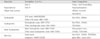
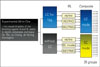 | Figure 1Bonding procedure and experimental grouping. LC; light cure, IRL; intermediate resin layer, SC; self cure, HI; hydrophilic, HO; hydrophobic, MO; mostly hydrophobic, f; filled
|
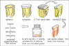 | Figure 2A schematic representation of the preparation of serial slabs from bonded composite, the application of nail varnish, immersion in the ammoniacal silver nitrate tracer.
|
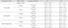
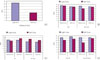 | Figure 3Bar charts showing the micro shear bond strength for adhesive only (A), with hydrophilic intermediate resin (B), with hydrophobic intermediate resin (C) and with most hydrophobic intermediate resin (D) according to the bonding protocols.
|
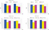 | Figure 4Bar charts compared to the micro shear bond strengths according to hydrophilicity of intermediate resin.
(A) no filled intermediate resin without cure of adhesive, (B) no filled intermediate resin after cure of adhesive, (C) filled intermediate resin without cure of adhesive, (D) filled intermediate resin after cure of adhesive.
|
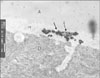
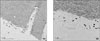 | Figure 5Transmission electron micrographs of the resin dentin interfaces after immersion in 50wt% silver nitrate. An experimental single step adhesive was bonded with light cure mode composite. A) × 3,400, B) × 7,100: Hybrid layer (H) was formed uniformly between dentin (D) and adhesive layer (A), and its thickness was approximately 2.0 µm. Nanoleakage, in the form of reticular patterns of silver deposits could be seen in the hybrid layer and along dentinal tubule after immersion with silver nitrate tracer.
|
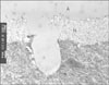
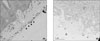 | Figure 6TEM micrographs of an experimental adhesive bonded with self cure mode composite. A) The resin dentin interface; The thickness of hybrid layer (H) was not evenly formed. Much of silver deposits were identified within and top of hybrid layer. Adhesive layer (A) was totally separated and silver deposits were infiltrated into adhesive layer (arrows). B) The resin composite interface; The interface was very irregular and worm eaten appearance. The interface of composite (C) side was hollowed with some adhesive and unidentified mass. Some of silver grains were shown along the interface in adhesive side.
|
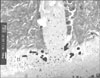
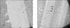 | Figure 7TEM micrographs of the resin dentin interfaces of an experimental adhesive with light cured composite after the hydrophilic intermediate resin layer (IRL) applied. A) IRL was directly applied on adhesive layer without curing. The hybrid layer was well formed and it was hard to identify any silver deposit. The thickness of IRL and adhesive layer was approximately 12 µm. B) IRL was applied after light curing of adhesive layer. Hybrid layer was also well formed and it was hard to identify any silver deposit. The thickness of IRL and adhesive layer was relatively thicker approximately 25 µm. There were definite demarcation line between adhesive layer and IRL (arrows).
|
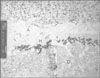
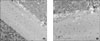 | Figure 8TEM micrographs of the resin dentin interfaces an experimental adhesive with self cured composite after the hydrophilic IRL applied. The IRL was directly applied on adhesive layer without curing. A) × 4,400, B) × 5,600: Hybrid layer was formed evenly and it was also hard to identify any silver deposit. The thickness of hybrid layer was approximately 10 µm.
|
 | Figure 9TEM micrographs of the resin dentin interfaces an experimental adhesive with light cured composite after the hydrophobic IRL applied (× 8,000). Collagen fibrils within the hybrid layer (H) appeared electron lucent as the undemineralized sections were not stained prior to TEM examination. Nanoleakage, in the form of reticular patterns of silver deposits could be seen in the hybrid layer and along dentinal tubule after immersion with silver nitrate tracer (arrows). Numerous isolated silver grains present throughout the hybrid layer and adhesive layer.
|
 | Figure 10TEM micrographs of the resin dentin interfaces an experimental adhesive with light cured composite after the most hydrophobic IRL applied (× 12,000). The hybrid layer was well formed and its thickness was approximately 2 µm. Collagen fibrils within the hybrid layer (H) clearly appeared electron lucent at this high magnification. The isolated silver grains were unevenly distributed in both the hybrid layer and lower side of dentinal tubule.
|
 | Figure 11TEM micrographs of the resin dentin interfaces an experimental adhesive with light cured composite after the most hydrophobic filled IRL applied (×10,000). Nanoleakage, in the form of reticular patterns of silver deposits could be seen in the hybrid layer (arrows). Numerous isolated silver grains were unevenly distributed in both the hybrid layer and dentinal tubule.
|
 | Figure 12TEM micrographs of the resin dentin interfaces an experimental adhesive with self cured composite after the most hydrophobic filled IRL applied (× 6,000). Nanoleakage, in the form of reticular patterns of silver deposits could be seen in the hybrid layer. Microfiller particles and silver grains could be seen within the intermediate resin layer (I). The adhesive layer (A) was relatively thin (approximately 3 µm) compared to the intermediate resin layer.
|
References
1. Tay FR, Pashley DH, Suh B, Carvalho R, Miller M. Single step, self etch adhesives behave as permeable membranes after polymerization. Part I. Bond strength and morphologic evidence. Am J Dent. 2004. 17:271–278.
2. Tay FR, Pashley DH, Garcia Godoy F, Yiu CK. Single step, self etch adhesives behave as permeable membranes after polymerization. Part II. Silver tracer penetration evidence. Am J Dent. 2004. 17:315–322.
3. Krämer N, Lohbauer U, Frankenberger R. Adhesive luting of indirect restorations. Am J Dent. 2000. 13(Spec No):60D–76D.
4. Rosenstiel SF, Land MF, Crispin BJ. Dental luting agents: a review of the current literature. J Prosthet Dent. 1998. 80:280–301.

5. Diaz Arnold AM, Vargas MA, Haselton DR. Current status of luting agents for fixed prosthodontics. J Prosthet Dent. 1999. 81:135–141.
6. Cheong C, King NM, Pashley DH, Ferrari M, Toledano M, Tay FR. Incompatibility of self etch adhesives with chemical/dual cured composites: two step vs one step systems. Oper Dent. 2003. 28:747–755.
7. Tay FR, Frankenberger R, Krejci I, Bouillaguet S, Pashley DH, Carvalho RM, Lai CN. Single bottle adhesives behave as permeable membranes after polymerization. I. In vivo evidence. J Dent. 2004. 32:611–621.
8. Chersoni S, Suppa P, Grandini S, Goracci C, Monticelli F, Yiu C, Huang C, Prati C, Breschi L, Ferrari M, Pashley DH, Tay FR. In vivo and in vitro permeability of one step self etch adhesives. J Dent Res. 2004. 83:459–464.
9. Sanares AM, Itthagarun A, King NM, Tay FR, Pashley DH. Adverse surface interactions between one bottle light cured adhesives and chemical cured composites. Dent Mater. 2001. 17:542–556.
10. Tay FR, Pashley DH. Aggressiveness of contemporary self etching systems. I: Depth of penetration beyond dentin smear layers. Dent Mater. 2001. 17:296–308.
11. Kubo S, Yokota H, Sata Y, Hayashi Y. Microleakage of cervical resin composite restoration. Modern trends in adhesive dentistry Proceedings of the adhesive dentistry forum '99 in Tsurumi. 1999. Yokohama, Japan: Kuraray Co. Ltd.;153–165.
12. Tay FR, Pashley DH, Yiu CK, Sanares AM, Wei SH. Factors contributing to the incompatibility between simplified step adhesives and chemically cured or dual cured composites. Part I. Single step self etching adhesive. J Adhes Dent. 2003. 5:27–40.
13. Tay FR, Suh BI, Pashley DH, Prati C, Chuang SF, Li F. Factors contributing to the incompatibility between simplified step adhesives and chemically cured or dual cured composites. Part II. Single bottle, total etch adhesive. J Adhes Dent. 2003. 5:91–105.
14. Tay FR, King NM, Suh BI, Pashley DH. Effect of delayed activation of light cured resin composites on bonding of all in one adhesives. J Adhes Dent. 2001. 3:207–225.
15. Tay FR, Pashley DH, Suh BI, Carvalho RM, Itthagarun A. Single step adhesives are permeable membranes. J Dent. 2002. 30:371–382.
16. Tay FR, Pashley DH, Peters MC. Adhesive permeability affects composite coupling to dentin treated with a self etch adhesive. Oper Dent. 2003. 28:610–621.
17. Moll K, Haller B. Effect of intrinsic and extrinsic moisture on bond strength to dentine. J Oral Rehabil. 2000. 27:150–165.
18. Pashley EL, Zhang Y, Lockwood PE, Rueggeberg FA, Pashley DH. Effects of HEMA on water evaporation from water HEMA mixtures. Dent Mater. 1998. 14:6–10.
19. Tay FR, Pashley DH. Water treeing - A potential mechanism for degradation of dentin adhesives. Am J Dent. 2003. 16:6–12.
20. Carrilho MRO, Carvalho RM, Tay FR, Pashley DH. Long term durability of resin dentin bond: a microtensile bond strength study. Am J Dent. in press.
21. Carrilho MRO, Carvalho RM, Tay FR, Pashley DH. Effects of storage media on mechanical properties of adhesive systems. Am J Dent, in press. Am J Dent. 2004. 17:104–108.
22. Carvalho RM, Pegoraro TA, Tay FR, Pegoraro LF, Silva NR, Pashley DH. Adhesive permeability affects coupling of resin cements that utilise self etching primers to dentine. J Dent. 2004. 32:55–65.
23. El Zohairy AA, De Gee AJ, Hassan FM, Feilzer AJ. The effect of adhesives with various degrees of hydrophilicity on resin ceramic bond durability. Dent Mater. 2004. 20:778–787.
24. Carrilho MR, Carvalho RM, Tay FR, Pashley DH. Effects of storage media on mechanical properties of adhesive systems. Am J Dent. 2004. 17:104–108.
25. Yiu CK, King NM, Pashley DH, Suh BI, Carvalho RM, Carrilho MR, et al. Effect of resin hydrophilicity and water storage on resin strength. Biomaterials. 2004. 25:5789–5796.
26. Yiu CK, King NM, Carrilho MR, Sauro S, Rueggeberg FA, Prati C, Carvalho RM, Pashley DH, Tay FR. Effect of resin hydrophilicity and temperature on water sorption of dental adhesive resins. Biomaterials. 2006. In press.
27. Ito S, Hashimoto M, Wadgaonkar B, Svizero N, Carvalho RM, Yiu C, et al. Effects of resin hydrophilicity on water sorption and changes in modulus of elasticity. Biomaterials. 2005. 26:6449–6459.
28. Yiu CK, Pashley EL, Hiraishi N, King NM, Goracci C, Ferrari M, Carvalho RM, Pashley DH, Tay FR. Solvent and water retention in dental adhesive blends after evaporation. Biomaterials. 2005. 26:6863–6872.
29. Prati C, Chersoni S, Acquaviva GL, Breschi L, Suppa P, Tay FR, Pashley DH. Permeability of marginal hybrid layers in composite restorations. Clin Oral Investig. 2005. 9:1–7.
30. King NM, Hiraishi N, Yiu CK, Pashley EL, Loushine RJ, Rueggeberg FA, Pashley DH, Tay FR. Effect of resin hydrophilicity on water vapour permeability of dental adhesive films. Eur J Oral Sci. 2005. 113:436–442.
31. Choi SM, Choi KK, Park SJ. Effect of the additional application of a resin layer on dentin bonding using single step adhesives. J Korean Acad Conserv Dent. 2006. in Press.
32. Tay FR, Pashley DH, Yoshiyama M. Two modes of nanoleakage expression in single step adhesives. J Dent Res. 2002. 81:472–476.
33. Chang YI, Choi KK, Park SJ. Compatibility between two step self etching adhesives and composites with different curing mode. J Korean Acad Conserv Dent. 2006. in Press.
34. Han CS, Choi KK, Choi GW. Influence of application/drying time of self etching primer on shear bond strength to dentin. Kyung Hee Dent J. 2001. 23:175–185.
35. Tanaka J, Ishikawa K, Yatani H, Yamashita A, Suzuki K. Correlation of dentin bond durability with water absorption of bonding layer. Dent Mater J. 1999. 18:11–18.
36. Burrow MF, Inokoshi S, Tagami J. Water sorption of several bonding resins. Am J Dent. 1999. 12:295–298.
37. Sano H, Yoshikawa T, Pereira PN, Kanemura N, Morigami M, Tagami J, Pashley DH. Long term durability of dentin bonds made with a self etching primer, in vivo. J Dent Res. 1999. 78:906–911.
38. Wang Y, Spencer P. Hybridization efficiency of the adhesive/dentin interface with wet bonding. J Dent Res. 2003. 82:141–145.
 XML Download