

 PDF
PDF ePub
ePub Citation
 Print
Print


I. INTRODUCTION
The ideal root canal sealer should present, among other physical/chemical properties, enough radiopacity to allow distinction from the adjacent anatomical structures1-4), such as bone and tooth5). Higginbotham6) was the first researcher to publish a study comparing the radiopacity of various endodontic sealers and gutta-percha cones used to fill root canals. Eliasson and Haasken7) established a comparison standard for radiopacity studies, using optical radiographic density measurements for impression materials and an equivalent thickness of aluminum capable of producing similar radiographic density. Beyer-Olsen and Orstavik1) included a reproducible comparison standard using an aluminum step wedge with 2 mm-increments to determine the radiopacity of several root canal sealers.
Root canal sealers may affect the periapical tissue when extruded. In such condition, they could cause not only degeneration of the tissue lying underneath the endodontic sealers, but could also delay wound healing. Therefore, root canal sealers should have good biocompatibility8).
Sealers could be classified according to their main constituents, such as zinc oxide-eugenol, calcium hydroxide, resins, glass ionomers, etc.9). The biocompatibility of a specific root canal sealer remains one of the principal considerations for selecting an appropriate sealer for a dental restoration10). From the literature, it would appear that the side effects of the use of various root canal sealers have been previously studied to some extent11-13). Epoxy resin-based sealers have been introduced in endodontic practice because of their favorable characteristics, such as adhesion to tooth structure, long working time, easey manipulation, and good sealing ability14). Several studies showed that polymerized resin-based sealers presented less cytotoxicity compared to zinc oxide-eugenol and calcium hydroxide-based sealers15,16). However, the cytotoxic effects of resin-based root canal sealers related to their radiopacity have not yet been studied.
The purpose of this study was to evaluate the radiopacity and cytotoxicity of various resin-based root canal sealers in comparison with zinc oxide-eugenol and calcium hydroxide-based root canal sealers. In addition, this study aimed to investigate the correlation between the radiopacity and cytotoxicity of them.
II. MATERIALS AND METHODS
Material preparation
Five root canal sealers were evaluated in this study: AH 26 (Dentsply De Trey Gmbh, Konstanz, Germany), EZ fill (EDS, Hackensack, NJ, USA), AD Seal (Meta-Biomed, Cheongju, Korea), ZOB Seal (Meta-Biomed, Cheongju, Korea) and Sealapex (SybronEndo, Glendora, CA, USA). The materials were prepared according to manufacturers' instructions. Ten specimens, 10 mm in diameter and 1 mm in thickness, were fabricated from each material tested. Metallic matrices were made and impressions were taken using a light-bodied silicone-based impression material (Silagum, DMG, Hamburg, Germany). Samples of the prepared sealers were then inserted into the impressions and stored in a moist chamber at 37℃, until complete set.
Radiographic evaluation
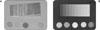
The specimens were placed on the occlusal x-ray film (Kodak Insight, Rochester, NY, USA) along with an aluminum (99.5% pure) step wedge with step heights ranging from 1 to 10 mm in increments of 1 mm (Figure 1A). A Kodak-2200 x-ray machine (Kodak, Rochester, NY, USA) operating at 70 kv, 10 mA, 18 pulses/s and with a focus-sensor distance of 30 cm was used. The digitized images (Figure 1B) were then imported into the Scion image software (Scion Corp. Frederick, MD, USA), where a tool was applied in order to identify the equal-density areas in the radiographic images.
Cell culture and inoculation
Human papilloma virus (HPV) 16-IPDL cells were obtained by transfecting normal human periodontal ligament (PDL) cells with pLXSN vector containing the E6/E7 open reading frames of HPV 16. This was done by the methods that have been previously described17). The IPDL cells were cultured in Dulbecco's modified Eagle's medium (DMEM; Biofluid, Rockville, MD, USA) supplemented with 10% fetal bovine serum (FBS) (Gibco, Carlsbad, CA, USA) containing 100 U/ml of both penicillin and streptomycin (Life Technologies, Rockville, MD, USA). The cultures were maintained at 37℃ in a humidified atmosphere of 5% CO2. Subsequently, single-cell suspensions of IPDL cells were seeded in 24-well tissue culture plates at 5 × 10⁴ cells per well, as determined by hemocytometry, in complete DMEM. The plates were incubated in a humidified atmosphere of 5% CO2 at 37℃ for 24 hours.
Cytotoxicity test
The five materials described previously were mixed and placed at the bottom of insert wells (n = 10) having a membrane pore diameter of 0.4 µm. After inserting the materials into the wells, the insert wells were exposed to ultraviolet (UV) light for 30 min and placed inside the culture wells; the cells were then incubated for 12, 24, 48, and 72 hours. All procedures were performed aseptically.
After exposure to the materials for 12, 24, 48, and 72 hours, the viable cells were detected using the MTT dye, which forms blue formazan crystals that are reduced by the mitochondrial dehydrogenase present in living cells. Briefly, 200 µl of MTT solution (2 mg/ml in phosphate-buffered saline [PBS]) was added to each well, and the wells were incubated for 4 hours. Subsequently, 200 µl of dimethyl sulfoxide (DMSO) was added to each well. The plates were then shaken until the crystals dissolved, and the solution in each well was transferred to a 96-well tissue culture plate. The reduced MTT was then measured spectrophotometrically at 540 nm in a dual beam microtiter plate reader. The cells that were incubated with the medium alone served as negative controls.
Statistical analysis
Statistical analysis was conducted by one-way analysis of variance. Tests of differences of the treatments were analyzed by Tukey test and a value of p < 0.05 was considered statistically significant. The comparison between radiopacity and cytotoxicity of resin-based root canal sealers were analyzed using two-way analysis of variance and Tukey test at a 95% significance level.
III. RESULTS
Radiographic evaluation
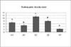
The results demonstrated that EZ fill presented the greatest radiopacity (p < 0.05) and were equivalent to 11.13 mm of aluminum. AH 26, AD Seal and ZOB Seal presented radiopacity values that were equivalent to 6.84, 4.82 and 7.85 mm of aluminum, respectively. Sealapex exhibited the lowest radiopacity (p < 0.05) and was equivalent to 2.22 mm of aluminum (Figure 2).
Cytotoxicity test
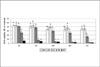
As shown in Figure 3, resin-based sealers (AH 26, EZ fill and AD Seal) showed statistically higher cell viabilities throughout all experimental times compared to other type of sealers. However, the cell viability of EZ fill was statistically lower than that of AD Seal in 24 and 48 and that of other two resin-based sealers in 72 hours. However, there was no correlation between the radiopacity and cytotoxicity of three resin-based root canals sealers (p > 0.05).
IV. DISCUSSION
Radiopacity is widely acknowledged as a desirable property of all intraoral materials, including root canal sealers18). Radiopacity of root canal sealers has been of particular significance for the evaluation of the quality of endodontic treatment, as well as being helpful in the assessment of possible voids in the obturation19). The ISO 6876/2001 establishes that root canal sealers should be at least as radiopaque as 3 mm thickness of aluminum2). Therefore, all sealers evaluated in this study presented the minimum radiopacity by this standard, except for Sealapex.
EZ fill contains zirconium and iron, which contributes for its greater radiopacity in relation to the other materials tested. Aoyagi et al.20) reported that radiopacity increased with the increase in radiopaque material content as well as the increase in atomic number of the element. We first hypothesized that the more radiopacity the resin-based root canal sealer shows, the more cytotoxicity would be presented. However, there was no statistical correlation between these two factors. This implies not only the content of radiopacifier but also other components contained in resin-based sealers contributed the cytotoxicity.
Sealapex presented radiopacity values slightly lower than those defined by the ISO. Kuga, et al.21) suggested the addition of iodoform to Sealapex, with the purpose of increasing the radiopacity. This addition did not affect the sealing properties or biocompatibility of materials.
In this present study, resin-based sealers showed lower cytotoxic effect compared to zinc oxideeugenol and calcium hydroxide-based sealers. Azar et al.15) reported that AH 26 was found to be highly toxic immediately after mixing, followed by a substantial decrease in cytotoxicity and this was due to formaldehyde. In the present study, however, pre-set sealers were applied to the cells and this might attribute to the low cytotoxicity.
In this study, zinc oxide-eugenol-based sealer, ZOB Seal, showed severe cytotoxicity throughout all experiment time. Many studies have investigated the biocompatibility of zinc oxide and eugenol-based root canal sealers22-26). Their cytotoxicity, however, is attributed to the eugenol, due to hydrophobic interactions with the cytoplasmic membrane27) or its effect on cell respiration28). Moreover, the zinc oxide contained in the root canal sealer is also dissociated when eugenol is released by hydrolysis29). Maseki et al.30) suggested that the toxicity may also be related to methyl salicylic acid, benzyl alcohol, zinc ions, rosin and other components released form the sealer. In a study by Valle et al.31), however, it was shown that the liquid to powder ratio is directly related to the toxicity of a zinc oxide and eugenol-based sealer. A higher ratio of liquid results in a more cytotoxic sealer. This implies that eugenol is by far the most toxic component.
Sealapex, a calcium hydroxide-based sealer containing neither eugenol nor formaldehyde, displayed the highest cytotoxicity. This seemed to be related to the method of applying the material to the cells. In the present study, the insert wells having a membrane pore diameter of 0.4 µm were used according to the method of Koulaouzidou et al.32). Schafer and Zandbiglari33) reported that calcium hydroxide-based root canal sealer has high solubility compared with resin or zinc oxide-eugenol. This contributed to release the ionic component from the material and subsequently lead to cytotoxicity. Further, these wells brought the tested materials in close proximity to the cells without interfering with the methods used for evaluating cell numbers, proliferation, and death.
V. CONCLUSIONS
Taken together, all sealers tested radiographically in this study presented the minimum radiopacity by this standard, except for Sealapex. In terms of cytotoxicity, resin-based sealers showed higher biocompatibility than other types of sealers. Further, EZ fill showed statistically lower cell viability compared to AD Seal in 24 and 48 hours and compared to other resin-based sealers in 72 hours. However, this result was not correlated with their radiopacity. In conclusion, these results indicate that resin-based root canal sealer is more biocompatible and has no disadvantage in terms of radiopacity.
 XML Download