

 PDF
PDF ePub
ePub Citation
 Print
Print


I. INTRODUCTION
Root canal preparation with nickel-titanium (NiTi) rotary instruments is easier and faster than with hand instruments1). NiTi instruments have two to three times higher elastic flexibility and a superior resistance to torsional fracture, compared with similar-sized conventional stainless steel instruments1-5). Also, rotary instrumentation facilitates the maintenance of the original canal shape and the position in space of the apical foramen6). Therefore those instruments can effectively produce a well-tapered root canal form sufficient for obturation with minimal risk of transporting the original canal, and are useful to the preparation of curved root canals1,3-5,7).
Various NiTi instrument systems have been introduced in the market; ProTaper (Dentsply Maillefer, Ballaigues, Switzerland), ProFile (Dentsply Maillefer, Ballaigues, Switzerland), Hero642 (Micromega, Besancon, France), K3 (SybronEndo, Glendora, France), etc. Generally, crown-down instrumentation sequence has been recommended for most of the rotary NiTi instruments, in which larger files precede smaller ones to progress further apically in turn8). The coronal preflaring might help to reduce the incidence of canal aberrations, and provide a better conical and tapered preparation that makes irrigation and spreader penetration easier9). Also, this technique is mandatory to reduce intracanal friction and thus to minimize the risk of instrument separation10).
Recently, new rotary NiTi instruments with different configuration and design have been marketed as Mtwo (VDW, Munich, Germany). The Mtwo instruments have a S-shaped cross-sectional design and a non-cutting safety tip. These instruments are characterized by a positive rake angle with two cutting edges. Moreover, Mtwo instruments have an increasing pitch length from the tip to the shaft. According to the manufacturer, the instruments should be used in a Single Length Technique and they claimed that it is easier than other method using variable working length. That means all files of the instrumentation sequence should be used to the full length of the root canal. This instrumentation technique is different from general method of rotary NiTi instruments10).
The Mtwo instruments cut dentine with their entire length and could advance all the way to the apex from the beginning of the sequence; consequently they are likely to be stressed more than other NiTi rotary instruments used in a crown-down progression then to make more canal aberrations and increase the risk of torsional fracture11).
Thus the aims of this study were to present a method of crown-down technique using Mtwo instruments and to compare the shaping ability and safety between single length technique and crown-down technique using Mtwo rotary instruments.
II. MATERIALS AND METHODS
1. Simulated root canals
Sixty simulated root canals in clear resin blocks (Endo Training Bloc; Dentsply Maillefer, Ballaigues, Switzerland) were used for this study. The root canals had a mean canal length of 17 mm and mean curvature angle of 40°
Aqueous methylene blue dye was injected into the canal to enhance the contrast of the preinstrumentation image. These resin blocks were scanned in a reproducible position with a scanner (Scanjet C8510A; Hewlett-Packard, California, USA), and then the data were stored in a personal computer.
2. Classification of groups
The simulated canals were divided into three groups according to the used instrument and the manner of using methods (Table 1). Each group had 20 specimens. Group MT resin blocks were instrumented with single length technique of Mtwo, group MC resin blocks with crown-down technique of Mtwo and group PT resin blocks with crown-down technique of ProTaper.
3. Preparation
All of the rotary NiTi files used in this study were operated by an electric motor (X-Smart; Dentsply Maillefer, Ballaigues, Switzerland) set at 250 r.p.m. and torque of 2.2 N without autoreverse function to count the frequencies of instrument binding. These setting values were within the range suggested at the manufacturer's operation manuals. The use of files was also in accordance with the manufacturer's recommendation.
During the preparation, canals were verified the patency with #10 K-Flexofile (Dentsply Maillefer, Ballaigues, Switzerland), then irrigated, and lubricated with RC-prep (Stone Pharmaceuticals, Philadelphia, USA) repeatedly after instrumentation. Files were wiped regularly with a sponge to remove resin debris. All files used in this study were new. The root canals were copiously irrigated with water dispensed through a 27-gauge needle (ENDO-EZE; Ultradent, South Jordan, USA) after using each instrument. After filing, apical gauging was verified with #25 K-Flexofile.
1) Group MT
The Mtwo file system is composed of .04 / #10, .05 / #15, .06 / #20 and .06 / #25 for basic sequence and .05 / #30, .04 / #35, .04 / #40 and .07 / #25 for further shaping.
In group MT, all Mtwo instruments were used to the full length of the canals according to the manufacturer's instructions of basic sequence using a gentle in-and-out motion. The instrument was used as following sequence (Table 2).
Once the instrument had negotiated to the end of the canal and had rotated freely, it was removed.
2) Group MC
The instrumentation sequence of group MC was designed through the preliminary test of preparation using simulated canal of resin block.
In group MC, Mtwo instruments were used in crown-down manner. Instruments were withdrawn when resistance was felt and changed to the next instrument. The instrument was used as following sequence (Table 2).
3) Group PT
ProTaper instruments were also used in crown-down manner according to the manufacturer's instructions using a gentle in-and-out motion (Table 2).
4. Image taking and superimposition
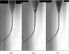
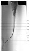
After the root canal preparation was completed, enlarged canals were filled with Vitapex (Neo Dental Chemical Products co., LTD, Tokyo, Japan) to enhance the contrast of the post-instrumentation image. Then the resin blocks were scanned again using the reproducible position. The pre- and post-instrumented canal images were superimposed on one another with the guide of attached label using Adobe Photoshop ver.7.0 (Adobe, San Jose, California, USA) (Figure 1). These superimposed images of 2736 × 944 pixel size were inspected on the TFT-LCD monitor (SyncMaster CX1565N; Samsung, Suwon, Korea) of 1024 × 768 screen resolutions. These images were used to evaluate the centering ratio and canal curvature (Figure 2).
5. Measurement and calibration
2) Centering ratio
Instrumented widths were measured linearly from the pre-instrumented point to the post-instrumented wall inwardly and outwardly. Measurements were taken horizontally at eight different levels (1, 2, 3, 4, 5, 6, 7 and 8 mm) from the level of apical foramen (Figure 3). These values were used to calculate the instrumented canal width and post-instrumented canal diameter14).
The centering ratio was computed to evaluate the movement of root canal's center after preparation. The ratio was calculated using the following method: the absolute value of net transportation divided by whole width of the post-instrumented canal. The centering ratio is a measure of the ability of an instrument to stay centered in the canal15-17).
3) Canal aberrations
Assessments were made according to the presence of various types of canal aberrations such as apical zip, elbow and ledge using the scanned post-instrumented images.
4) Weight loss
The weights of resin blocks were measured before and after preparation using ANALYTICAL Plus Balance (AP110; OHAUS, Pine Brook, NJ, USA). Loss of the weight was calculated by subtracting the post-instrumented weight from the pre-instrumented one.
5) Instrumentation time
Instrumentation time was recorded in seconds by one assistant. The recorded time excluded the time for irrigation, changing instruments and patency.
6. Statistical analysis
Statistical analysis of the collected data was performed with SPSS version 10.0 (SPSS Inc., Chicago, IL, USA). Mean scores for each group were statistically compared using ANOVA and Duncan's Multiple Range Test for post-hoc comparison. The Fisher's exact test was done for evaluation of canal aberration and instrument binding. Differences revealed in the data were designated as significant at p < 0.05.
III. RESULTS
1. Reduction of canal curvature
There was no significant difference in the reduction of root canal curvature (degree) between groups (p > 0.05) (Table 3).
3. Canal aberrations
The group MT showed more elbows than group MC and group PT (p < 0.05) (Table 5). Ledge was not occurred in any group.
4. Weight loss
There was no significant difference in weight loss of resin block after preparation between groups (p > 0.05) (Table 6).
5. Instrumentation time
In instrumentation time, the group MT and group MC required shorter preparation time than group PT (p < 0.05). Despite of the frequency of instrument change, group MC showed no significant difference with group MT (Table 7).
6. Frequency of instrument binding
The group MC had the least frequency of file binding and the group PT had the most (p < 0.05) (Table 8).
7. Instrument distortion
In group MT, there were 2 files of distortion. These 2 files were .04 / #10. But, there was no significant difference between three groups (p > 0.05) (Table 9). Instrument fracture was not occurred in any group.
IV. DISCUSSION
Recent advances in the field of endodontics have led to the use of nickel titanium rotary instruments in dental practice. The NiTi rotary instrumentation has several advantages compared to stainless steel hand instrumentation1). During the last a few years the number of users has increased significantly and many new systems have been on the market. When a new system is introduced, clinicians should keep the manufacturer's recommendation and the basic principle of canal preparation for successful clinical application.
The Mtwo system is one of the NiTi instruments currently available. The manufacturer's recommended instruction for usage of Mtwo rotary instruments is different from general method of other NiTi rotary system. The Single Length Technique suggested by manufacturer seems to induce more stress to the instruments and more screwing.
The purposes of this study were to compare the shaping effect and safety between single length technique and crown-down technique and to suggest more safe method using Mtwo rotary instrument.
The ProTaper system is an active file system which has the flute of blade type and Mtwo system has same type of flute. The cross-section of Mtwo file is S-shape while that of ProTaper is triangular18). There were a few reports about Mtwo rotary11,18,19), however, several researches have been studied for the safety and the efficiency of crown-down technique using ProTaper system20-22). The study using mathematic models have demonstrated that ProTaper instruments work longer in a super elastic phase than instruments with a U-file design, allowing for high performance and less risk20). According to Calberson et al.21), ProTaper instruments performed acceptable tapered preparations in all canal types. Iqbal et al.22) reported that ProTaper system is able to optimally enlarge root canal with minimal transportation and loss of working length in vitro study compared with ProFile system. Thus, the ProTaper instruments served as controls in order to ensure the results obtained from Mtwo rotary instrument with crown-down technique under identical experimental conditions.
There was no significant difference in the reduction of root canal curvature (degree), centering ratio of each level and weight loss of resin block after preparation between three experimental groups.
From the view point of centering ratio, the smaller the ratio, the better the instrument remained centered in the canal15-17). In the present study, the centering ratios at the levels of 1 mm and 5 mm indicate that all experimental groups deviated severely. Several researches23-25) about ProTaper reported the large centering ratio and transportation tendency after completion of root canal preparation at apical portion.
Mtwo system also showed similar apical deviation and coronal straightening regardless of using manner. It might be resulted from morphologic similarity of file flute and cutting tendency to ProTaper. The change in using sequence of Mtwo instrument makes no difference in qualitative and quantitative outcomes of root canal preparation. From the viewpoint of apical centering, regardless of using manner of Mtwo instruments, it is demanded to use carefully especially in apical canal portion as well as ProTaper.
One of the main problems inherent in the rotary preparation by clinicians is the instrument fracture9). In this study, there was no statistical difference in instrument distortion that occurred only group MT, which can be interpreted due to using new files in each canal26) and the adoption of torque control motors for the experiment26,27). The fact that none of the Mtwo files fractured after enlargement of canal may be due to the increasing pitch length from the tip to the shaft of these instruments8). As already reported, a varying pitch length along the working part of the instrument reduces the screw-in tendency of the file to minimize the risk of instrument fracture28).
However, some care should be taken using the Mtwo files with single length technique because of the occurrence of permanent deformations of the group MT. Although no statistical difference between groups, two Mtwo instruments were deformed permanently in group MT. Consequently, if the files are used repeatedly, instruments could be fractured.
In instrumentation time, the group MT and group MC required shorter preparation time than group PT. Despite of the frequency of instrument change, the group MC showed no significant difference from group MT. Although the preparation by group MC needed three more files than group MT, group MC needed no more preparation time. It seemed that enlarged coronal canal by crown-down usage of Mtwo file would facilitate the access to apical canal and this could shorten the time lapse of each step in group MC than that of group MT. In other studies14,24) to compare ProTaper and other instruments, instrumentation time with ProTaper was shorter than other systems under identical experimental conditions. But in this study, the Mtwo file showed shorter preparation time than ProTaper, regardless of using method of Mtwo file. This result could not be compared to other study because there was no report to compare these two systems.
The group MT showed more elbows than group MC and group PT. There was no ledge formation in any group. The aberration (elbow) might leave a significant portion of un-instrumented canal wall which would be difficult to obturate29). In ProTaper system, tooth structure can be removed excessively when this instrument is left in the canal too long with its active design21). It is therefore of utmost importance to follow the manufacturer's instructions and not to leave the ProTaper to prepare the root canal for longer than 1 second or not to use the ProTaper too much when reaching the desired working length14). This precaution would also be applied in Mtwo instruments of group MT in which showed more aberrations.
These aberrations might be produced by the using manner of single length technique. It is assumed that group MT might make apical aberrations similar to many aberrations in apical shaping of step-back technique resulted from insufficient coronal flaring. The coronal preflaring might help to reduce the incidence of canal aberrations in group MC. And these aberrations can be diminished more through several pre-clinical trial uses especially with newly introduced instrument.
The group MC had the least frequency of file binding and the group PT had the most. The Mtwo system was used, in group MT, according to the sequence suggested because each of these instruments is claimed to be able to enlarge the whole canal. The resin debris attached to file flute was shown in entire file length in group MT but a portion of flute in group MC. The Mtwo instruments of group MT cut dentine with their entire length and could advance all the way to the apex from the beginning of the sequence, consequently they are likely to be stressed more than the instruments used in a crown-down progression11). In crown-down technique, the use of larger and high taper instruments initially can lead to an unobstructed apical access for the smaller files30). This principle can make safe usage of NiTi file with minimal binding or screwing and most of NiTi systems follow this manner of sequence. In the preliminary investigation for modification of the recommended sequence, the .04 / #10 file was revealed to work less in the simulated canal because the favorable coronal flaring attained already with .07 / #25 file and .06 / #20 file before working length measurement. Then in MC group, the .04 / #10 file was omitted. These modified sequences seemed to lessen the binding frequency.
The crown-down technique of instruments reduced contact area and reduced engagement zone. Torque was correlated with apically directed force and contact areas31). As binding increases, torsional stress by locking file tip into canal wall seems to introduce the high torsional stress and exceeds the elastic limit that leads to plastic deformation and ductile fracture32). Likewise, group MT seems to introduce more torsional stresses as the binding increases. According to Sattapan et al32), the taper lock effect is believed to occur when instruments with the same taper, but varying tip diameters in step-back method, are wedged into a canal, possibly leading to torsional overloading of the instrument tip. The Mtwo file in group MT showed much more taper lock effect than group MC, especially in the instant of reaching at working length.
The single length technique of group MT seems to be similar to the apical shaping procedure from initial apical file to master apical file in step-back method. The Quantec file, one of the systems early introduced, used a sequence of same working length through the apical preparation stage of gradually increasing taper with same apical tip size33). This system is not used in these days because it seems to have high risk of fracture from taper lock.
The frequency of binding, in this study, might be influenced by operator's proficiency. The operator proficiency, undoubtedly, is a significant factor in reducing fracture incidence of NiTi rotary instruments, probably by avoiding the screwing31). The untrained operator probably exerts excess apical pressure on instrument in the canal. Consequently, the instruments locks into the canal and are subjected to high level torque. Instrument deformation occurs and the incidence of separation increase significantly. The preclinical training is a prerequisite to increase the reliability of the instrumentation technique as well as keeping the technical guidelines strictly.
The simulated canals in clear resin blocks were used to control the experimental conditions of this study that might influence the shaping abilities of the instruments. The simulated canals in resin blocks used in this study does not permit the action of the instruments in root canals of real teeth because of differences in the surface texture, hardness and cross-section. However, resin blocks allow a direct comparison of the shaping ability of different instruments and studies on extracted teeth fully confirmed observations made on resin blocks3,5). As use of simulated canals in resin blocks would have more binding of instruments, the study of with the use of real teeth will be necessary.
From the present results of this study, we can suggest that crown-down technique using Mtwo rotary file not only showed similar shaping ability to single length technique recommended by manufacturer but also seemed to have more safety.
V. CONCLUSION
Under the limited condition of this study, crown-down technique using Mtwo rotary file not only showed similar shaping ability to single length technique recommended manufacturer but also seems to have more safety to minimize the binding or screwing which can induce the torsional fracture.


 XML Download