

 PDF
PDF ePub
ePub Citation
 Print
Print


Abstract
C-shaped canals are known to present a complex canal anatomy with numerous fins connecting individual canals, thus requiring supplementary effort to accomplish a successful root canal treatment. This study examined the frequency of the C-shaped mandibular second molars and interrelation between the clinical records and radiographs to recognize them treated in the Department of Conservative Dentistry of the Chosun University Dental Hospital during a six-year period (1998 - 2004). This study reviewed the clinical records of 227 patients who underwent root canal treatment of the mandibular second molars. After opening the chamber, those cases with C-shaped orifices in the pulpal floor were selected, and the C-shaped root canal types were classified according to Melton's criteria. Three experienced dentists evaluated the radiographs of the C-shaped mandibular second molar on a viewer using a magnifying glass in order to determine if the root apex was fused or separated, the distal root canal was either centered or mesial shifted in the distal root, and if there was bilateral symmetry in a panorama. In conclusion, there is a high frequency of C-shaped mandibular second molars in Koreans. Simultaneous interpretation of the root shape and distal root canal using the preoperative, working length and post-treatment radiographs is important for diagnosing a C-shaped mandibular second molar.
Figures and Tables
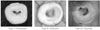
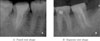
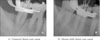
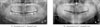
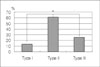
Figure 5
Frequency of the C-shaped root canal types.
*: Significantly different at p < 0.01 (χ2 test)
References
1. Cooke HG, Cox FL. C-shaped canal configurations in mandibular molars. J Am Dent Assoc. 1979. 99:836–839.

2. Yang ZP, Yang SF, Lin YL. C-shaped root canals in mandibular first molars in Chinese population. Endod Dent Traumatol. 1988. 4:160–163.
3. Weine FS. Members of Arizona Endodontic Association. The C-shaped mandibular second molar: incidence and other considerations. J Endod. 1998. 24:372–375.
4. Dankner E, Friedman S, Stabholz A. Bilateral C shape configuration in maxillary first molars. J Endod. 1990. 16:601–603.
5. De Moor RJG. C-shaped root canal configuration in maxillary first molars. Int Endod J. 2002. 35:200–208.
6. Bolger WL, Schindier WG. A mandibular first molar with a C-shaped root configuration. J Endod. 1988. 14:515–519.
7. Rice RT, Gilbert BO. An unusual canal configuration in a mandibular first molar. J Endod. 1987. 13:515–515.
8. Melton DC, Krell KV, Fuller MW. Anatomical and histological features of C-shaped canals in mandibular second molars. J Endod. 1991. 17:384–388.
9. Fan B, Cheung GS, Fan M, Gutmann JL, Bian Z. C-shaped Canal System in Mandibular Second Molars: Part I-Anatomical Features. J Endod. 2004. 30:899–903.
10. Lambrianidis T, Lyroudia K, Pandelidou O, Nicolaou A. Evaluation of periapical radiographs in the recognition of C-shaped mandibular second molars. Int Endod J. 2001. 34:458–462.
11. Walid N. The Use of Two Pluggers for the Obturation of an Uncommon C-Shaped Canal. J Endod. 2000. 26:422–424.
12. Liewehr FR, Kulild JC, Primack PD. Obturation of a C-Shaped Canal Using an Improved Method of Warm Lateral Condensation. J Endod. 1993. 19:474–477.
13. Ricucci D, Pascon EA, Langeland K. Long-Term Follow-Up on C-Shaped Mandibular Molars. J Endod. 1996. 22:185–187.
14. Manning SA. Root canal anatomy of mandibular second molars. Part II C-shaped canals. Int Endod J. 1990. 23:40–45.
15. Chai WL, Thong YL. Cross-sectional Morphology and Minimum Canal Wall Widths in C-shaped Roots of Mandibular Molars. J Endod. 2004. 30:509–512.
16. Lyroudia K, et al. 3D Reconstruction of Two C-Shape Mandibular Molars. J Endod. 1997. 23:101–104.
17. Fan B, Cheung GS, Fan M, Gutmann JL, Fan W. C-shaped canal system in mandibular second molars: Part II-Radiographic Features. J Endod. 2004. 30:904–908.
18. Al-Fouzan KS. C-shaped root canals in mandibular second molars in a Saudi Arabian population. Int Endod J. 2002. 35:499–504.
19. Haddad GY, Nehme WB, Ounsi HF. Diagnosis, Classification, and Frequency of C-Shaped Canals in Mandibular Second Molar in the Lebanese Population. J Endod. 1999. 25:268–271.
20. Seo MS, Park DS. C-shaped root canals of mandibular second molars in a Korean population: clinical observation and in vitro analysis. Int Endod J. 2004. 37:139–144.
21. Sabala CL, Benenati FW, Neas BR. Bilateral root or root canal aberrations in a dental school patient population. J Endod. 1994. 20:38–42.
22. Martin H, Cunningham WT, Norris JP, Cotton WR. Ultrasonic vs. hand filing of dentin: a quantitative study. Oral Surg. 1980. 49:79–84.
23. Langeland K, Liao K, Pascon EA. Working-saving devices in endodontics: efficiency of sonic and ultrasonic techniques. J Endod. 1985. 11:499–510.
24. Reynolds MA, Madison S, Walton RE, Krell KV, Rittman BR. An intro histological comparison of the step-back, sonic and ultrasonic instrumentation techniques in small, curved root canals. J Endod. 1987. 13:307–314.
 XML Download