

 PDF
PDF ePub
ePub Citation
 Print
Print


Abstract
The fracture toughness test is believed as a clinically relevant method for assessing the fracture resistance of the dentinal restoratives. The objectives of this study were to measure the fracture toughness (K1C) and microtensile bond strength of dentin-resin composite interface and compare their relationship for their use in evaluation of the integrity of the dentin-resin bond.
A minimum of six short-rod specimens for fracture toughness test and fifteen specimens for microtensile bond strength test was fabricated for each group of materials used. After all specimens storing for 24 hours in distilled water at 37℃, they were tensile-loaded with an EZ tester universal testing machin. Statistical analysis was performed using ANOVA and Tukey's test at the 95% confidence level, Pearson's coefficient was used to verify the correlation between the mean of fracture toughness and microtensile bond strength. FE-SEM was employed on fractured surface to describe the crack propagation.
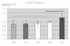
Fracture toughness value of Clearfil SE Bond (SE) was the highest, followed by Adper Single Bond 2 (SB), OptiBond Solo (OB), ONE-STEP PLUS (OS), ScotchBond Multi-purpose (SM) and there was significant difference between SE and other 4 groups (p < 0.05). There were, however, no significant difference among SB, OB, OS, SM (p > 0.05). Microtensile bond strength of SE was the highest, followed by SB, OB, SM, OS and OS only showed significant lower value (p < 0.05). There was no correlation between fracture toughness and microtensile bond strength values. FE-SEM examination revealed that dentin bonding agent showed different film thickness and different failure pattern according to the film thickness.
From the limited results of this study, it was noted that there was statistically no correlation between K1C and µTBS. We can conclude that for obtaining the reliability of bond strength test of dentin bonding agent, we must pay more attention to the test procedure and its profound scrutiny.
Figures and Tables

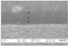
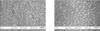
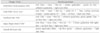
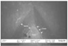 | Figure 6The resin surface of fractured K1C specimen of SM group (× 50).
It showed that bevel formed by spacer and crack occurred along the bevel.
C: Composite resin, CAD: Composite adhered to dentin
|
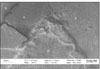
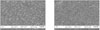
 | Figure 7The resin surface of fractured K1C specimen of OB group (× 500).
The arrows showed that crack formed along the bevel move into the adhesive surface (see the black arrows).
|
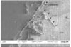
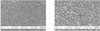
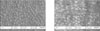
 | Figure 8The dentin surface of fractured K1C specimen of SB group (× 250).
It revealed that the failure occurred between resin-infiltrated layer and adhesive layer.
At the tip of the bonded surface(white arrow), failure occurred mainly within the adhesive resin layer.
D: dentin, AR: Adhesive resin layer
|
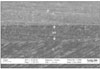 | Figure 9The cross-sectional SEM photograph of bonded surface of SM group (× 1,000).
The thickness of adhesive layer was 40 ~ 55 µm and dentinal tubules were running oblique direction.
The adhesive layer was relatively thick and uniform.
|
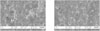
 | Figure 10The cross-sectional SEM photograph of bonded surface of SB group (× 3,000).
The thickness of adhesive layer was 14 ~ 25 µm and the dentinal tubules were filled with resin tags.
Some of the dentinal tubules were empty, some of the resin tags had many branches.
RT: Resin tag, ID: Intertubular dentin, PD: Peritubular dentin
|
 | Figure 11The cross-sectional SEM photograph of bonded surface of SE group (× 1,000).
The thickness of adhesive layer was 50 ~ 60 µm and it was the thickest adhesive resin layer among the experimental groups.
Most of dentinal tubules were filled with resin tags.
|
 | Figure 12The resin surface (left) and dentin surface (right) of fractured SM group (× 2,000).
Bond failure occurred predominantly between the resin-infiltrated layer and adhesive resin.
The orifices of dentinal tubules were not seen clearly and irregular adhesive resin layer was showed at the dentin surface of fractured specimen.
|
 | Figure 13The resin surface (left) and dentin surface (right) of fractured OS group (× 2,000).
Many broken resin tags were seen on the both specimens,
the fracture of the resin tags mainly occurred at the upper part of the resin-infiltrated layer.
RT: resin tag, DT: dentinal tubule
|
 | Figure 14The resin surface (left) and dentin surface (right) of fractured OB group (× 2,000).
On the resin surface, many resin tags and adhesive resin layer was seen partially.
The dentin surface of the fractured specimen showed relatively smooth appearance and many dentinal tubules filled with resin tags. The fracture of resin tags were occurred at the orifices of the dentinal tubules.
AR: adhesive resin layer, RT: resin tag
|
 | Figure 15The resin surface (left) and dentin surface (right) of fractured SB group (× 2,000).
Relatively short broken resin tags were seen on the resin side.
Irregular adhesive resin layer and dentinal tubules obstructed with resin tag or covered with adhesive resin were seen on the dentin side of the fractured specimen.
|
 | Figure 16The resin surface (left) and dentin surface (right) of fractured SE group (× 2,000).
Resin tags and dentinal tubules were seen rarely.
Both sides showed very irregular appearance and bond failure occurred predominantly within the adhesive resin layer.
|


References
1. Nakabayashi N. Resin reinforced dentin due to infiltration of monomers into the dentin at the adhesive interface. J Dent Mater. 1982. 1:78–81.
2. Van Meerbeek B, Inokoshi S, Braem M, Lambrechts P, Vanherle G. Morphological aspects of the resin-dentin interdiffusion zone with different dentin adhesive systems. J Dent Res. 1992. 71:1530–1540.

3. Eick JD, Robinson SJ, Chappell RP, Cobb CM, Spencer P. The dentinal surface ; its influence on dentinal adhesion. Part III. Quintessence Int. 1993. 24:571–582.
4. Van Meerbeek B, Dhem A, Goret-Nicaise M, Braem M, Lambrechts P, Vanherle G. Comparative SEM and TEM examination of the ultrastructure of the resin-dentin interdiffusion zone. J Dent Res. 1993. 72:495–501.
5. Tay FR, Gwinnett AJ, Pang KM, Wei SHY. Structural evidence of a sealed tissue interface with a total-etch wet-bonding technique in vivo. J Dent Res. 1994. 73:629–636.
6. Van Meerbeek B, Mohrbacher H, Celis JP, Roos JR, Braem M, Lambrechts P, Vanherle G. Chemical characterization of the resin-dentin interface by micro-Raman spectroscopy. J Dent Res. 1993. 72:1423–1428.
7. Eick JD, Miller RG, Robinson SJ, Bowles CQ, Gutshall PL, Chappelow CC. Quantitative analysis of the dentin adhesive interface by Auger spectroscopy. J Dent Res. 1996. 75:1027–1033.
8. Kruger MB. Raman mapping of the dentin/adhesive interface. Appl Spectrosc. 1996. 50:1500–1504.
9. Burke FJT, McCaughey AD. The four generations of dentin bonding. Am J Dent. 1995. 8:88–92.
10. Pashley DH, Sano H, Ciucchi B, Yoshiyama M, Carvalho RM. Adhesion testing of dentin bonding agents : a reviews. Dent Mater. 1995. 11:117–125.
11. Swift EJ Jr, Perdigão J, Heymann HO. Bonding to enamel and dentin : a brief history and state of the art. Quintessence Int. 1995. 26:95–110.
12. Heymann HO, Sturdevant JR, Bayne S, Wilder AD, Sluder TB, Brunson WD. Examining tooth flexure effects on cervical restorations : A two-year clinical study. J Am Dent Assoc. 1991. 122:41–47.
13. Duke ES, Robbins JW, Schwartz RS, Summitt JB, Conn LJ. Clinical and interfacial laboratory evaluation of a bonding agent in cervical abrasions. Am J Dent. 1994. 7:307–311.
14. van Dijken JWV. Clinical evaluation of four dentin bonding agents in class V abrasion lesions : a four-year follow-up. Dent mater. 1994. 10:319–324.
15. Van Meerbeek B, Peumans M, Verschueren M, Gladys S, Braem M, Lambrechts P, Vanherle G. Clinical status of ten dentin adhesive systems. J Dent Res. 1994. 73:1690–1702.
16. Yamaguchi R, Powers JM, Dennison JB. Parameters affecting in vitro bond strength of composites to enamel and dentin. Dent Mater. 1989. 5:153–156.
17. Chappell RP, Eick JD. Shear bond strength and scanning electron microscopic observation of current dentinal adhesives. Quintessence Int. 1994. 25:359–368.
18. Sorensen JA, Dixit NV. In vitro shear bond strength of dentin adhesives. Int J Prosthodont. 1991. 4:117–125.
19. Söderholm KJ. Correlation of in vivo and in vitro performance of adhesive restorative materials : a report of the ASC MD156 Task Group on Test Methods for the Adhesion of Restorative Materials. Dent Mater. 1991. 7:74–83.
20. Retief DH. Standardizing laboratory adhesion tests. Am J Dent. 1991. 4:231–236.
21. Tyas MJ. Guest editorial: reliability and validity in dental materials testing. J Dent Res. 1991. 70:1471.
22. Jendresen MD, Allen EP, Bayne SC, Hansson TL, Klooster J, Preston JD. Report of the committee on scientific investigation of the American Academy of Restorative Dentistry. J Prosthet Dent. 1992. 68:137–190.
23. van Noort R, Noroozi S, Howard IC, Cardew G. A critique of bond strength measurements. J Dent. 1989. 17:61–67.
24. van Noort R, Cardew GE, Howard IC, Noroozi S. The effect of local interfacial geometry on the measurement of the tensile bond strength to dentin. J Dent Res. 1991. 70:889–893.
25. Wang CT, Pilliar RM. Bond cement bonding-interfacial fracture toughness determination. Clin Mater. 1989. 4:135–153.
26. Charalambides PG, Cao HC, Lund J, Evans AG. Development of a test method for measuring the mixed mode fracture resistance of bimaterial interfaces. Mech Mater. 1990. 8:269–283.
27. Tam LE, Pilliar RM. Fracture toughness of dentin/resin-composite adhesive interfaces. J Dent Res. 1993. 72:953–959.
28. Lin CP, Douglas WH. Failure mechanisms at the human dentin-resin interface: A fracture mechanics approach. J Biomech. 1994. 27:1037–1047.
29. Hertzberg RW. Deformation and fracture mechanics of engineering materials. 1983. 2nd ed. New York: J. Wiley.
30. Kelly JR. Perspectives on strength. Dent Mater. 1995. 11(2):103–110.
31. Beaumont PWR, Young RJ. Slow crack growth in acrylic bone cement. J Biomed Mater Res. 1975. 9:423–439.
32. Mecholsky JJ. Fracture mechanics principles. Dent Mater. 1995. 11(2):111–112.
33. Griffith AA. The phenomena of rupture and flow in solids. Philos Trans R Soc Lond A. 1920. 221:163–198.
34. Ferracane JL, Antonio RC, Matsumoto H. Variables affecting the fracture toughness of dental composites. J Dent Res. 1987. 66(6):1140–1145.
35. Lloyd CH, Mitchell L. The fracture toughness of tooth coloured restorative materials. J Oral Rehabil. 1984. 11:257–272.
36. Ferracane JL, Berge HX. Fracture toughness of experimental dental composites aged in ethanol. J Dent Res. 1995. 74(7):1418–1423.
37. Young RJ, Beaumont PWR. Failure of brittle polymers by slow crack growth. Part 2. Failure processes in a silica particle-filled epoxy resin composite. J Mater Sci. 1975. 10:1343–1350.
38. Kim KH, Park JH, Imai Y, Kishi T. Microfracture mechanisms of dental resin composites containing spherically-shaped filler particles. J Dent Res. 1994. 73(2):499–504.
39. Lloyd CH, Adamson M. The development of fracture toughness and fracture strength in posterior restorative materials. Dent Mater. 1987. 3:225–231.
40. Goldman M. Fracture properties of composite and glass ionomer dental restorative materials. J Biomed Mater Res. 1985. 19:771–783.
41. de Groot R, Van Elst HC, Peters MC. Fracture-mechanics parameters of the composite-enamel bond. J Dent Res. 1990. 69:31–35.
42. Barker LM. A simplified method for measuring plane strain fracture toughness. Eng Fract Mech. 1977. 9:361–369.
43. Bubsey RT, Munz D, Pierce WS, Shannon JL. Compliance calibration of the short rod chevron-notch specimen for fracture toughness testing of brittle material. Int J Frac. 1982. 125–133.
44. Van Meerbeek B, De Munck J, Yoshida Y, Inoue S, Vargas M, Vijay P, Van Landuyt K, Lambrechts P, Vanherle G. Adhesion to Enamel and Dentin : Current Status and Future Challenges. Oper Dent. 2003. 28(3):215–235.
45. Chung SM, Yap AU, Tsai KT, Yap FL. Elastic modulus of resin-based dental restorative materials : a microindentation approach. J Biomed Mater Res B Appl Biomater. 2005. 72(2):246–253.
46. Fujishima A, Ferracane JL. Comparison of four modes of fracture toughness testing for dental composites. Dent Mater. 1996. 12:38–43.
47. Van Meerbeek B, De Munck J, Yoshida Y, Shirai K, Inoue S, Shiatani H, Lambrechts P. Chemical bonding potential of adhesive materials to hydroxyapatite. J Dent Res. 2003. 82(Special Issue):
48. Tam LE, Khoshand S, Pilliar RM. Fracture resistance of dentin-composite interfaces using different adhesive resin layers. J Dent. 2001. 29:217–225.
49. Ai H, Nagai M. Effect of the adhesive layer thickness on the fracture toughness of dental adhesive resin. Dent Mater J. 2000. 19(2):153–163.
50. Rueggeberg FA, Margeson DH. The effect of oxygen inhibition on the unfilled/filled composite system. J Dent Res. 1990. 69:1652–1658.
51. Finger WJ, Lee KS, Podszun W. Monomers with low oxygen inhibition as enamel/dentin adhesives. Dent Mater. 1996. 12:256–261.
52. Miyazaki M, Ando S, Hinoura K, Onose H, Moore BK. Influence of filler addition to bonding agents on shear bond strength to bovine dentin. Dent Mater. 1995. 11:234–238.
53. Nunes MF, Swift EJ, Perdigăo J. Effects of adhesive composition on microtensile bond strength to human dentin. Am J Dent. 2001. 14(6):340–343.
54. Frankenberger R, Lopes M, Perdigão J, Ambrose WW, Rosa BT. The use of flowable composites as filled adhesives. Dent Mater. 2002. 18(3):227–238.
55. Retief DH, Wendt SL, Bradley EL. Effect of adhesive thickness on the shear bond strength of Scotchbond 2/Silux to dentin. Am J Dent. 1989. 2(6):341–344.
56. Leloup G, D'hoore W, Bouter D, Degrange M, Vreven J. Meta-analytical Review of Factors Involves in Dentin Adherence. J Dent Res. 2001. 80(7):1605–1614.
57. Tantbirojn D, Cheng YS, Versluis A, Hodges JS, Douglas WH. Nominal Shear or Fracture Mechanics in the Assessment of Composite-Dentin Adhesion? J Dent Res. 2000. 79(1):41–48.
 XML Download