

 PDF
PDF ePub
ePub Citation
 Print
Print


I. Introduction
The aim of root canal instrumentation is to create a tapered shape with adequate volume to allow effective irrigation and obturation1). The ideal preparation of the root canal is a funnel shaped form with the smallest diameter at the apex and the widest diameter at the orifice2). However, traditional stainless steel (SS) instruments often failed in achieving these objectives, especially when in severely curved canals3-5). The bigger the size of the SS instrument, the more it tends to straighten up canal curvature in narrow and curved canal. It causes some problems like ledge, zip, perforation, and canal transportation which lead endodontic failure6).
In order to eliminate some of the shortcomings of these SS instruments, rotary nickel-titanium (Ni-Ti) instruments have been developed. These instruments are two to three times more flexible than SS instruments and also markedly superior to SS instruments in terms of angular deflection and maximum torque to failure7,8). These new instruments have been found to be better than SS instruments in maintaining the original anatomy, shape and position of the apical foramen9). According to Glosson et al.10) these instruments produce a better-centered and rounder canal preparation in comparison with SS instruments.
Many rotary Ni-Ti file systems have been introduced to the market. Most of these Ni-Ti file systems - e.g. ProFile® (Dentsply Maillefer, Ballaigues, Switzerland), K3™ (SybronEndo, Glendora, France), Hero642® (Micromega, Besancon, France) - have a constant tapered shaft design, while they have various rake angles and radial lands respectively11-13). Recently introduced ProTaper® system (Dentsply Maillefer, Ballaigues, Switzerland) which sales in two way of rotary and manual type, has been found to incorporate instruments of progressive multitaper design with sharp cutting blades. And it was designed to provide the fewest number of instruments that would afford improved flexibility, efficiency and safety14). It was claimed that ProTaper® system provides a continuous tapered preparation of the root canal, without significant transportation of the original position15).
However, after the ProTaper® system was introduced, the possibility of more or less severe canal transportation produced by active cutting action was discussed. Peter et al.16) showed that the ProTaper® system tends to transport canals slightly larger than other file systems with a passive cutting action by micro CT evaluation of shaped canal studies. Lee and colleagues17) demonstrated that ProTaper® files remove too much canal structure and cause severe canal transportation than other files by the study using resin blocks.
The purpose of this study was to investigate that a certain file steps in the ProTaper® system influences on canal transportation mainly through analysis of root canal instrumentation step by step.
II. Materials and Methods
Twenty simulated root canals in clear resin blocks (Endo Training Bloc; Dentsply Maillefer, Ballaigues, Switzerland) were used for this study. The root canals had a mean canal length of 17 mm and mean curvature of 40° as determined by Schneider's method18).
The canals were divided into two groups according to instrument. Group R-resin blocks were instrumented with rotary ProTaper® and group M-resin blocks were instrumented with manual ProTaper®.
1. Image taking of pre-operative blocks
The resin blocks were scanned in a reproducible position with a scanner (Scanjet® C8510A, Hewlett-Packard, California, USA). Aqueous red ink was injected into the canals to enhance the image contrast.
2. Instrumentation
Before the Ni-Ti files were used, the canals were explored with stainless steel #10 hand K-files until the tip was visible at the apical foramen. The working lengths were established to be 1 mm short from the apical foramen. Canals were prepared with a crown-down method according to the recommended sequences of the manufacturers. RC-Prep® (Stone Pharmaceuticals, Philadelphia, USA) was used as a lubricant. During the procedures, all simulated canals were verified the patency with #10 hand K-file. The root canals were irrigated after each instrument use with normal saline dispensed through a 27-gauge needle (ENDO-EZE®, Ultradent, South Jordan, Utah, USA). Preparation sequence summarized at Table 1.
2-1. Rotary ProTaper® instrumentation
The electric motor (Tecnika®, ATR, Pistola, Italy) set at a speed of 300 rpm and torque of 30 (Tecnika motor setting value) in a 16 : 1 reduction handpiece was used. Rotary ProTaper® instruments were withdrawn when resistance was felt and changed for the next instrument. According to the manufacturer's recommendation, the measured length was 13 mm when light resistance was felt using ISO stainless steel #15 K-file. Shaping file no.1 (S1) was used first to 13 mm length. Then auxiliary shaping file (SX) was used to same length, followed by shaping files no.1 (S1) and no.2 (S2) to the working length for the shaping of the coronal two thirds of the canal. The apical one third was finished by using finishing files no.1 (F1) and no.2 (F2) sequentially to the working length.
2-2. Manual ProTaper® instrumentation
According to the manufacturer's recommendation, manual ProTaper® was inserted with clockwise rotation and gentle inward pressure until it started to bind against the canal wall. Next, the file was withdrawn with counterclockwise rotation regularly to remove debris and check flutes. Instrumentation sequence was the same in rotary ProTaper® instrumentation.
3. Image taking of post-operative blocks
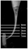
After each file step preparation, aqueous methylene blue solution was injected into the enlarged canals. Acquired resin blocks were scanned again in a reproducible position. Original canal image and the image after using each file step were superimposed for calculation of centering ratio. The image after using each file step and image after using previous file step were superimposed for calculation of the amount of deviation. All of these superimposed images were assessed on a 17 inch TFT-LCD monitor (Sync Master® CX701N, Samsung, Suwon, Korea) using Adobe® Photoshop software and were observed at a magnification of 156 times. Measurements were taken horizontally at five different levels (1, 2, 3, 4 and 5 mm) from the level of apical foramen (Figure 1).
4. Measurement techniques
4-1. Centering ratio
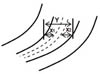
Centering ratio was calculated using the following method: the absolute value of net transportation divided by whole width of the post-instrumented canal. Generally, centering ratio is calculated by comparing pre-and post-instrumented images after finishing the root canal preparation (Figure 2). But at this study, centering ratio was calculated after using each file; O-S1 file step, O-S2 file step, O-F1 file step and O-F2 file step.
4-2. Amount of deviation (The absolute value of net transportation)
Net transportation was determined from the discrepancy between outward and inward-instrumented width (Figure 2). At this study, the amount of deviation was calculated with the superimposed images using each file step image and previous file step image; O-S1 file step, S1-S2 file step, S2-F1 file step and F1-F2 file step.
III. Results
1. Centering ratio of each step
In rotary ProTaper® instrumentation group, the centering ratio of each step at all levels were not significantly different (p > 0.05).
In manual ProTaper® instrumentation group, there were no significant differences in all levels except of 1 mm (p > 0.05). At the level of 1 mm, the O-F2 file step showed the largest value, followed by O-F1 file step, O-S2 file step and O-S1 file step. It was significantly different between O-F2 file step and O-S1 file step (p < 0.05, Table 2).
2. Amount of deviation of each step
In rotary ProTaper® instrumentation group, amount of deviation of each step at all levels were not significantly different (p > 0.05).
In manual ProTaper® instrumentation group, there were no significant differences in all levels except of 1 mm (p > 0.05). At the level of 1 mm, the F1-F2 file step showed largest value, followed by S2-F1 file step, O-S1 file step and S1-S2 file step. F1-F2 file step was significantly different from results of other steps (p < 0.05, Table 3).
IV. Discussion
Recently introduced ProTaper® system represents a new generation of Ni-Ti instruments currently available. The basic series of ProTaper® files comprise six instruments, three shaping and three finishing files. The shaping files have a progressive taper sequence (increasing from tip to coronal) whereas the finishing files show a decreasing taper profile. It is claimed that the progressive taper sequence should enhance the flexibility of the files in the middle and at the tip region and that the decreasing taper sequence should enhance the strength of the files19). The manufacturer claims that these files are specially designed to instrument difficult, highly calcified, and severely curved root canals20).
The manual ProTaper® system that was launched recently has served a dual purpose. On the one hand it has introduced nickel-titanium to the clinician, who had previously wished to try the method but did not feel comfortable making the quantum leap to rotary NiTi system and on the other, it has been aimed as additional armamentarium for the clinician using rotary NiTi system, faced with more delicate, complex preparation or acute canal curvatures in apical regions21). Manual ProTaper® system has two advantages compared to rotary ProTaper®. First, it can be used in abrupt curvature with prebent instrument when pathway established by traditional hand files. Second, instrument separation is low due to good tactile feedback22).
Several researches have studied for the safety and the efficiency of ProTaper® system. The study using mathematic models have demonstrated that ProTaper® instruments work longer in a super elastic phase than do instruments with a U-file design, allowing for high performance and less risk23). According to Calberson et al.24), ProTaper® instruments performed acceptable tapered preparations in all canal types. Iqbal et al.25) reported that ProTaper® system is able to optimally enlarge root canal with minimal transportation and loss of working length in in vitro study compared with ProFile® system.
On the other hand, recently published study26) demonstrated that varying degrees of canal straightening and transportation towards the outer aspect of the curvature were evident when curved canal enlarged with ProTaper® instruments. Schäfer et al.27) reported that ProTaper® tended to transport towards the outer aspect of the canal curve. Peters et al.16) reported that ProTaper® design may also increase the incidence of procedural errors and overall canal transportation whilst the modified cutting flute of ProTaper® instruments may reduce friction and consequently torque.
Based on the results of this study, in rotary ProTaper® instrumentation group, centering ratio and the amount of deviation of each file step were not significantly different. To the contrary, in manual ProTaper® instrumentation group, F2 file step at apical 1 mm level had significantly large centering ratio and the amount of deviation.
Consequently, F2 file step of manual ProTaper® tended to transport the apical part of the canals than that of rotary ProTaper®. One possible explanation for this result may be related uneven rotational speed and torque, because clinicians use manual ProTaper® at their convenience.
Finishing files are greater in diameter than other Ni-Ti files at the same level of the root canal because of progressively different parabolic tapers28). This results in thicker and stiffer instruments. And they cause high lateral forces in curved canals. These restoring forces attempt to return the file to its original shape and act on the outer side on the canal wall during preparation, result in canal straightening and ledging. Therefore, manufacturer recommended that clinicians take the finishing files to the estimated length only once and remove them as soon as possible for proper shaping and to prevent canal aberrations28).
Previous studies27,28) comparing rotary ProTaper® system with other rotary Ni-Ti system have been reported about greater canal transportation tendency of rotary ProTaper® system. In the present study, no statistically significant differences were observed in canal transportation tendency of each file step of rotary ProTaper® system. But F2 file step of manual ProTaper® system showed greater canal transportation tendency at apical 1 mm level. Consequently, it is expected that clinicians can use rotary ProTaper® more accurately than manual ProTaper®.
V. Conclusion
In manual ProTaper® instrumentation group, F2 file step had significantly large centering ratio and the amount of deviation at the level of 1 mm (p < 0.05).
Under the condition of this study, F2 file step of manual ProTaper® tended to transport the apical part of the canals than that of rotary ProTaper®.


 XML Download