

 PDF
PDF ePub
ePub Citation
 Print
Print


Abstract
Competition will usually develop between the opposing walls as the restorative resin shrinks during polymerization. Magnitude of this phenomenon may be depended upon cavity configuration and volume.
The purpose of this sturdy was to evaluate the effect of cavity configuration and volume on microleakage of composite resin restoration that has margins on the enamel site only.
The labial enamel of forty bovine teeth was ground using a model trimmer to expose a flat enamel surface. Four groups with cylindrical cavities were defined, according to volume and configuration factor (Depth × Diameter / C-factor) - Group I: 1.5 mm × 2.0 mm / 4.0, Group II: 1.5 mm × 6.0 mm / 2.0, Group III: 2.0 mm × 1.72 mm / 5.62, Group IV: 2.0 mm × 5.23 mm / 2.54.
After treating with fifth-generation one-bottle adhesive - BC Plus™ (Vericom, AnYang, Korea), cavities were bulk filled with microhybrid composite resin - Denfill™ (Vericom). Teeth were stored in distilled water for one day at room temperature and were finished and polished with Sof-Lex system. Specimens were thermocycled 500 times between 5℃ and 55℃ for 30 second at each temperature.
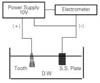
Teeth were isolated with two layers of nail varnish except the restoration surface and 1 mm surrounding margins. Electrical conductivity (µA) was recorded in distilled water by electrochemical method. Microleakage scores were compared and analyzed using two-way ANOVA at 95% level.
The results were as follows:
1. Small cavity volume showed lower microleakage score than large one, however, there was no statistically significant difference.
2. There was no relationship between cavity configuration and microleakage.
Factors of cavity configuration and volume did not affect on microleakage of resin restorations with enamel margins only.
Figures and Tables


References
1. Dentin bonding systems: an update. Council on Dental Materials, Instruments, and Equipment. J Am Dent Assoc. 1987. 114(1):91–95.
2. Nakabayashi N, Kosima K, Masuhara E. Promotion of adhesion by infiltration of monomer into tooth substrates. J Biomed Mater Res. 1982. 16:265–273.
3. Bowen RL. Properties of a silica-reinforced polymer for dental restoration. J Am Dent Assoc. 1963. 66:57–64.
4. Labella R, Lambrechts P, Van Meerbeek B, Vanherle G. Polymerization shrinkage and elasticity of flowable composite and filling adhesive. Dent Mater. 1999. 15:128–137.

5. Brännström M. Communication between the oral cavity and the dental pulp associated with restorative treatment. Oper Dent. 1984. 9:57–68.
6. Jörgensen KD, Asmussen E, Shimokobe H. Enamel damage by contracting restorative resin. Scand J Dent Res. 1975. 83:120–122.
7. Sheth JJ, Fuller JL, Jensen ME. Cuspal deformation and fracture resistance of teeth with dentin adhesives and composites. J Prosthet Dent. 1988. 60(5):560–569.
8. Choi KK, Ryu GJ, Choi SM, Lee MJ, Park SJ, Ferracane JL. Effects of cavity configuration on composite restoration. Oper Dent. 2004. 29(4):462–469.
9. Davidson CL, de Gee AJ. Relaxation of polymerization contraction stressed by flow in dental composite. J Dent Res. 1984. 63:146–148.
10. Feilzer AJ, de Gee AJ, Davidson CL. Quantitative determination of stress reduction by flow in composite restoration. Dent Mater. 1990. 6:167–171.
11. de la Macorra JC, Gomez-Fernandez S. Quantification of the configuration factor in Class I and II cavities and simulated cervical erosions. Eur J Prosthodont Restor Dent. 1996. 4(1):29–33.
12. Miguel A, de la Maccora JC. A predictive formula of the contraction stress in restorative and luting materials attending to free and adhesive surface, volume and deformation. Dent Mater. 2001. 17:241–246.
13. Ferracane JL, Mitchem JC. Relationship between composite contraction stress and leakage in class V cavities. Am J Dent. 2003. 16:239–243.
14. Pashley DH, Ciucchi B, Sano H, Horner JA. Permeability of dentin to adhesive agents. Quintessence Int. 1993. 24(9):618–631.
15. Van Meerbeek B, Inokoshi S, Braem M, Lambrechts P, Vanherle G. Morphological aspects of the resin-dentin interdiffusion zone with different dentin adhesive systems. J Dent Res. 1992. 71(8):1530–1540.
16. Van Meerbeek B, Peumans M, Verschueren M, Gladys S, Braem M, Lambrechts P, Vanherle G. Clinical status of ten dentin adhesive systems. J Dent Res. 1994. 73(11):1690–1702.
17. Gladys S, Van Meerbeek B, Lambrechts P, Vanherle G. Microleakage of adhesive restorative materials. Am J Dent. 2001. 14(3):170–176.
18. Hannig M, Friedrichs C. Comparative in vivo and in vitro investigation of interfacial bond variability. Oper Dent. 2001. 26(1):3–11.
19. Eick JD, Welch FH. Dentin adhesives-do they protect the dentin from acid etching? Quintessence Int. 1986. 17(9):533–544.
20. Johnston WM, Leung RL, Fan PL. A mathematical model for post-irradiation hardening of photoactivated composite resins. Dent Mater. 1985. 1(5):191–194.
21. Iwami Y, Yamamoto H, Ebisu S. A new electrical method for detecting marginal leakage of in vitro resin restorations. J Dent. 2000. 28(4):241–247.
22. Mattison GD, von Fraunhofer JA. Electrochemical microleakage study of endodontic sealer/cements. Oral Surg Oral Med Oral Pathol. 1983. 55(4):402–407.
23. Nakano Y. A new electrical testing method on marginal leakage of composite resin restorations. Japan J conserv Dent. 1985. 8(4):1183–1198.
24. Delivanis PD, Chapman KA. Comparison and reliability of techniques for measuring leakage and marginal penetration. Oral Surg Oral Med Oral Pathol. 1982. 53(4):410–416.
25. Buonocore MG. A simple method of increasing the adhesion of acrylic filling materials to enamel surfaces. J Dent Res. 1955. 34(6):849–853.
26. Gwinnett AJ, Matsui A. The physical relationship between enamel and adhesive. Arch Oral Biol. 1967. 12(12):1615–1620.
27. Retief DH. Effect of conditioning the enamel surface with phosphoric acid. J Dent Res. 1973. 52(2):333–341.
28. Silverstone LM. Fissure sealants. Laboratory studies. Caries Res. 1974. 8(1):2–26.
29. Gwinnett AJ. Histologic changes in human enamel following treatment with acidic adhesive conditioning agents. Arch Oral Biol. 1971. 16(7):731–738.
30. Eick JD, Robinson SJ, Cobb CM, Chappell RP, Spencer P. The dentinal surface: its influence on dentinal adhesion. Quintessence Int. 1992. 23(1):43–51.
31. Eick JD, Gwinnett AJ, Pashley DH, Robinson SJ. Current concepts on adhesion to dentin. Crit Rev Oral Biol Med. 1997. 8(3):306–335.
32. Swift EJ Jr, Perdigao J, Heymann HO. Bonding to enamel and dentin: a brief history and state of the art. Quintessence Int. 1995. 26(2):95–110.
33. Bastos PA, Retief DH, Bradley EL, Denys FR. Effect of etch duration on the shear bond strength of a microfilm composite resin to enamel. Am J Dent. 1988. 1(4):151–157.
34. Barkmeier WW, Shaffer SE, Gwinnett AJ. Effects of 15 vs 60 second enamel acid conditioning on adhesion and morphology. Oper Dent. 1986. 11(3):111–116.
35. Mitchem JC. The use and abuse of aesthetic materials in posterior teeth. Int Dent J. 1988. 38(2):119–125.
36. Frankenberger R, Kramer N, Petschelt A. Long-term effect of dentin primers on enamel bond strength and marginal adaptation. Oper Dent. 2000. 25(1):11–19.
37. Franco EB, Gonzaga Lopes L, Lia Mondelli RF, da Silva e Souza MH Jr, Pereira Lauris JR. Effect of the cavity configuration factor on marginal microleakage of esthetic restoration materials. Am J Dent. 2003. 16(3):211–214.
38. Mallmann A, Zovico F, Soares M, Placido E, Ferrari M, Cardoso PE. Microtensile dentin bond strength of self-etching and single-bottle adhesive system in different cavity configurations. J Adhes Dent. 2003. 5(2):121–127.
39. Armstrong SR, Keller JC, Boyer DB. The influence of water storage and C-factor on dentin-resin composite microtensile bond strength and pathway utilizing a filled and unfilled adhesive. Dent Mater. 2001. 17(3):268–276.
40. Barnes DM, Thompson VP, Blank LW, McDonald NJ. Microleakage of Class 5 composite resin restorations: a comparison between in vivo and in vitro. Oper Dent. 1993. 18(6):237–245.
41. Abdalla AI, Davidson CL. Comparison of the marginal integrity of in vivo and in vitro Class II composite restorations. J Dent. 1993. 21(3):158–162.
42. Ferrari M, Yamamoto K, Vichi A, Finger WJ. Clinical and laboratory evaluation of adhesive restorative systems. Am J Dent. 1994. 7(4):217–219.
43. Momoi Y, Iwase H, Nakano Y, Kohno A, Asanuma A, Yanagisawa K. Gradual increases in marginal leakage of resin composite restorations with thermal stress. J Dent Res. 1990. 69(10):1659–1663.
44. Kemp-Scholte CM, Davidson CL. Complete marginal seal of Class V resin composite restorations effected by increased flexibility. J Dent Res. 1990. 69(6):1240–1243.
 XML Download