

 PDF
PDF ePub
ePub Citation
 Print
Print


Abstract
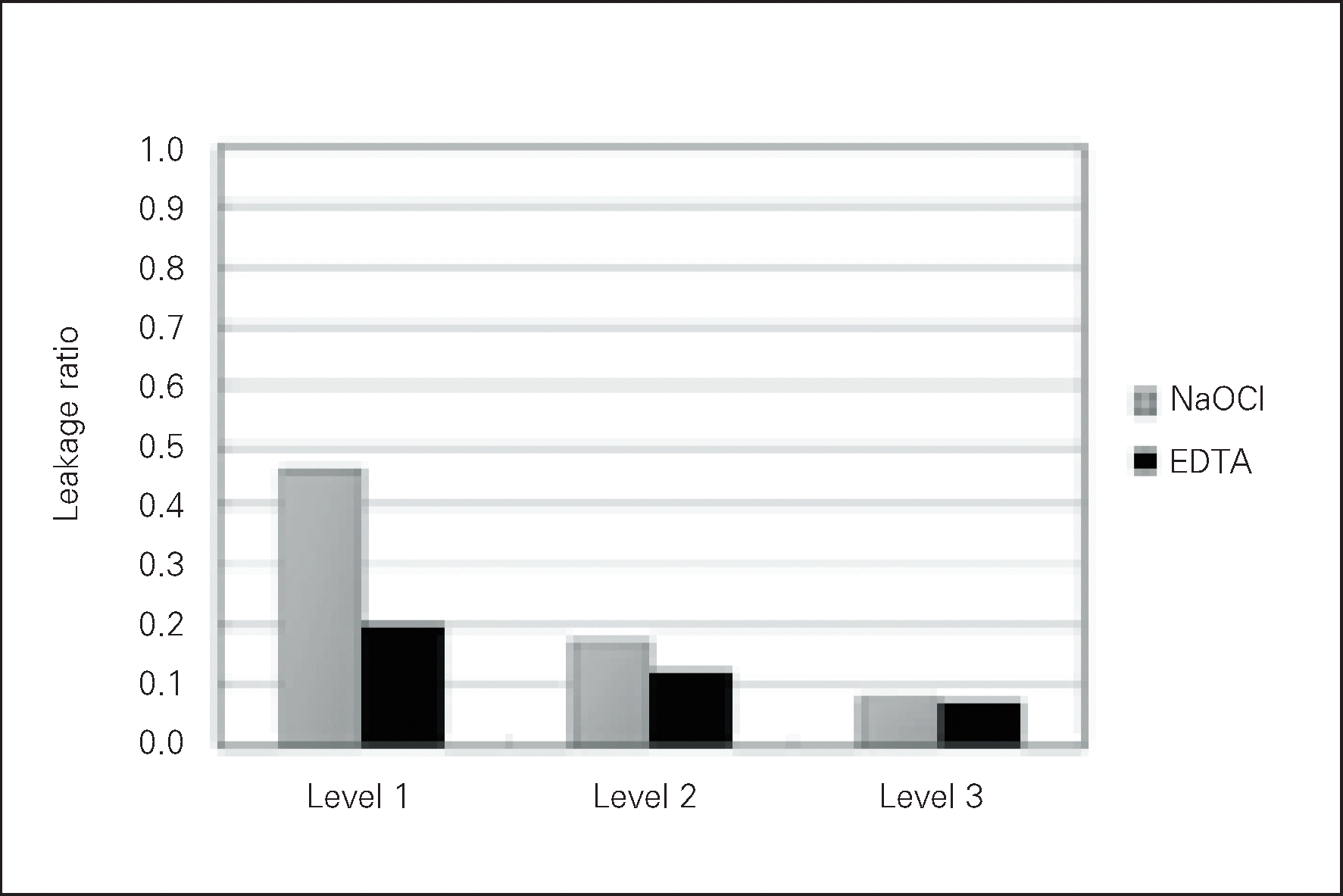
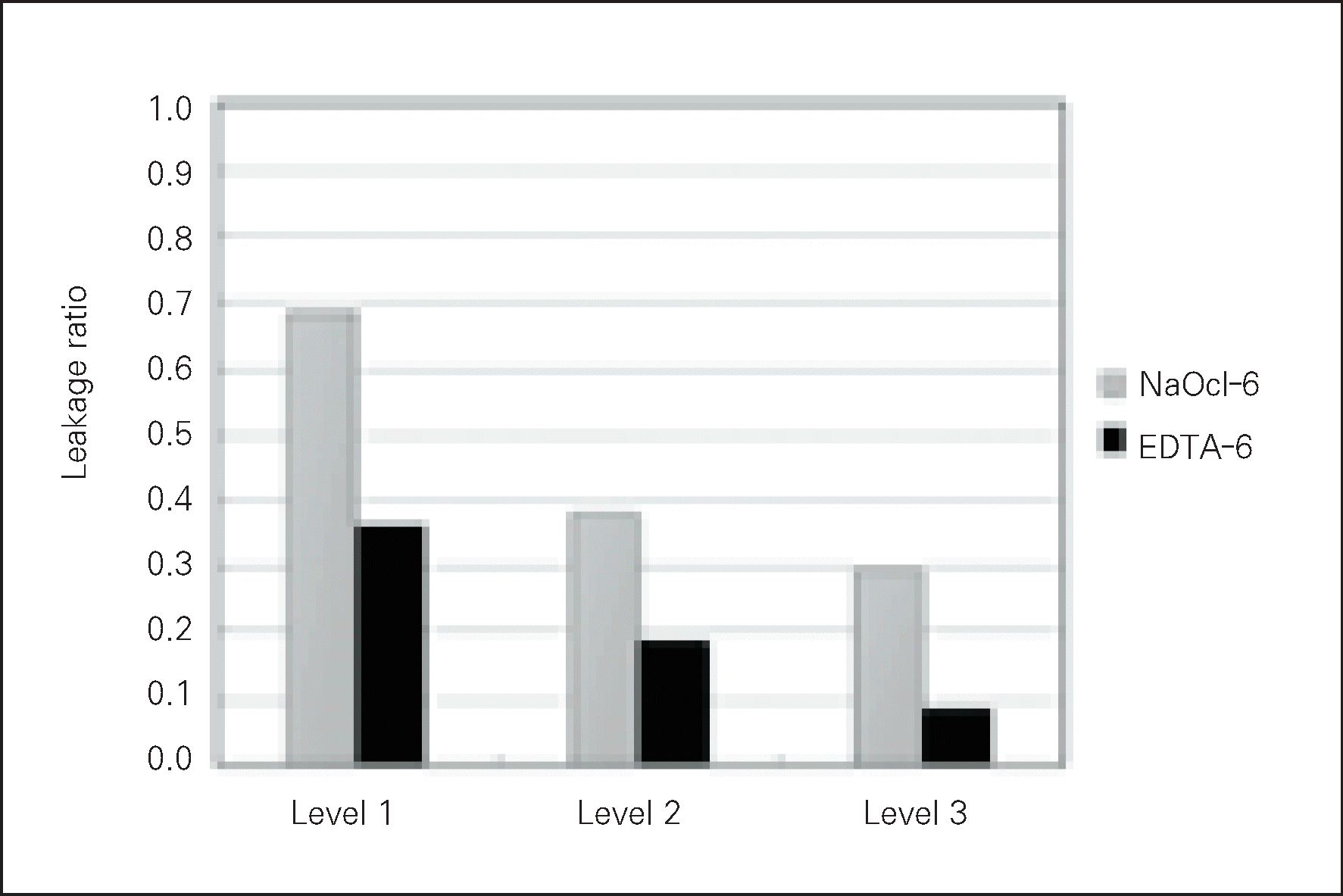
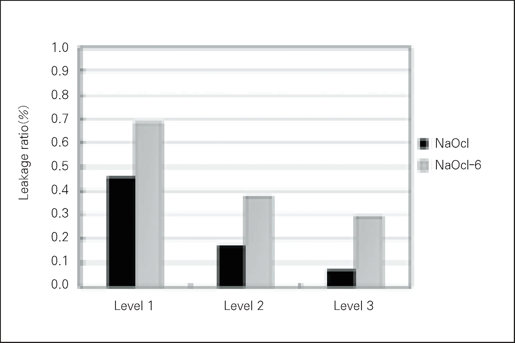
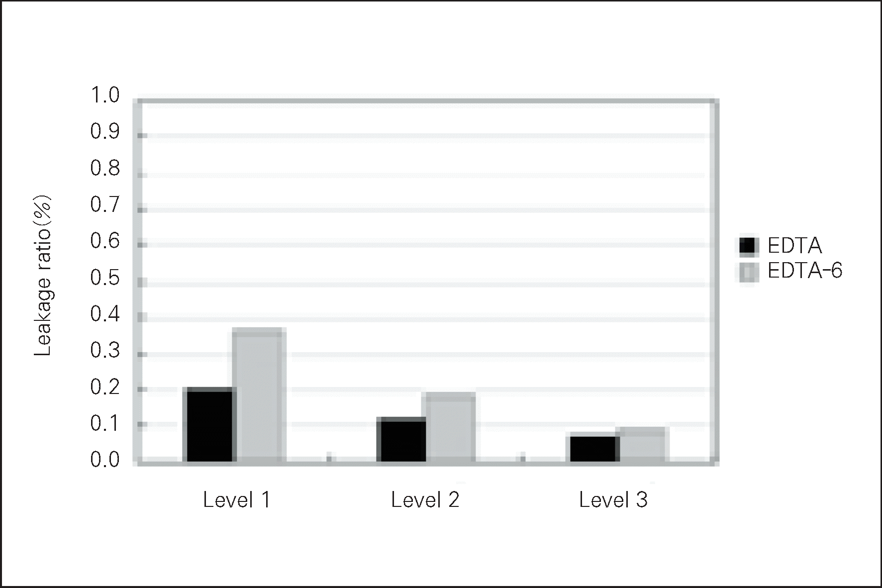
The mean leakage ratio was decreased cervically.
NaOCl group showed higher mean leakage ratio than EDTA group at each level. But there was significant difference at level 1 only (p < 0.05).
NaOCl-6 group showed higher mean leakage ratio than EDTA-6 group at each level. But there was significant difference at level 1 only (p < 0.05).
NaOCl-6 group showed higher mean leakage ratio than NaOCl group at each level. But there was significant difference at level 1 only (p < 0.05).
EDTA-6 group showed higher mean leakage ratio than EDTA group at each level. But there was no significant difference.
In NaOCl group and NaOCl-6 group, scanning electron micrographs of tooth sections generally covered with smear layer. In EDTA group and EDTA-6 group, tooth sections showing the penetration of sealers to opened dentinal tubules. The results suggest that removal of smear layer was effective to reduce the apical microleakage of the root canal.
참고문헌
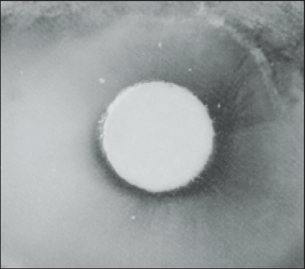
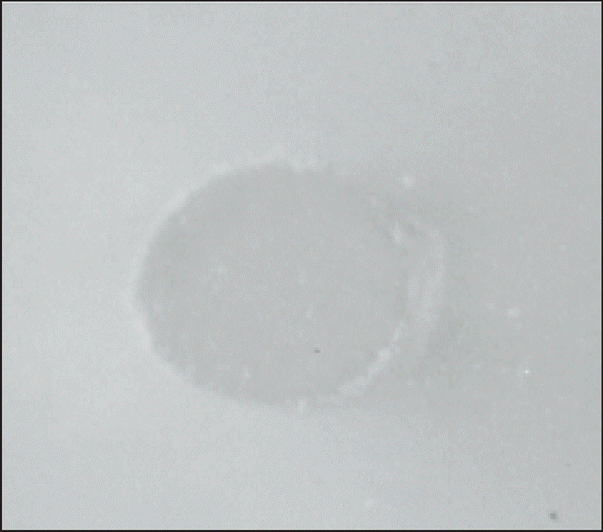
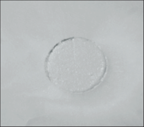
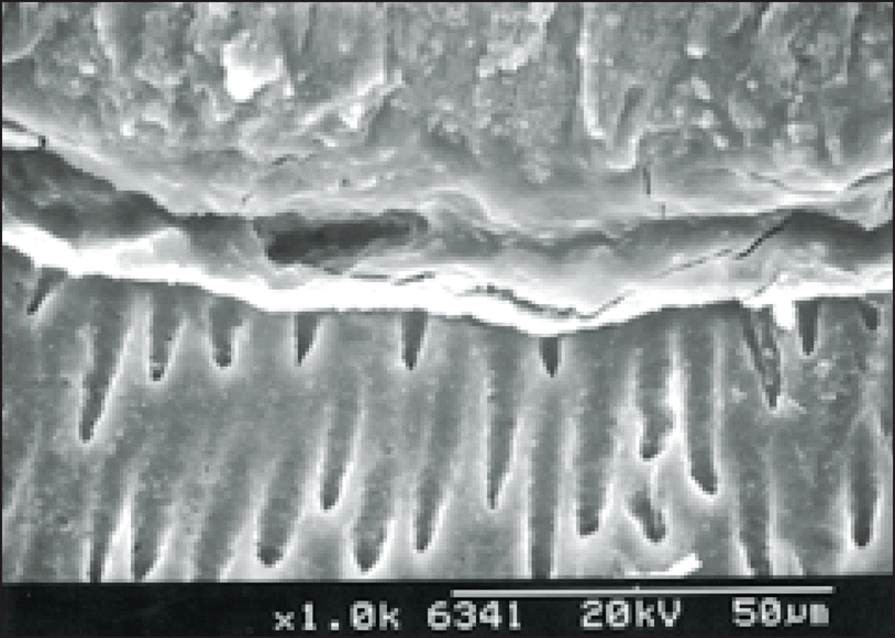
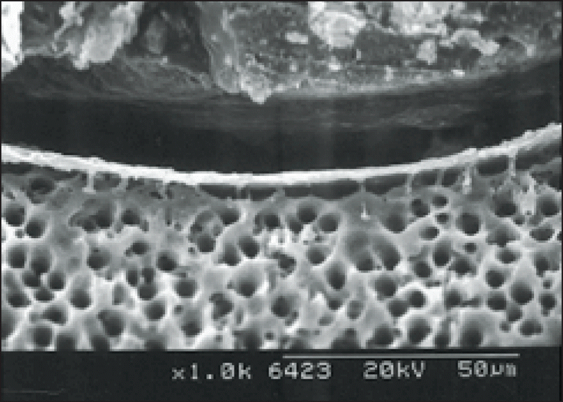 | Figure 17.Photograph of the NaOCl group Level 1 (× 1000); Scanning electron micrographs of tooth sections showing the gaps between gutta-percha and the surface of root canals. Surface generally covered with smear layer. Gaps were occuring between the smear layer and the gutta-percha. |
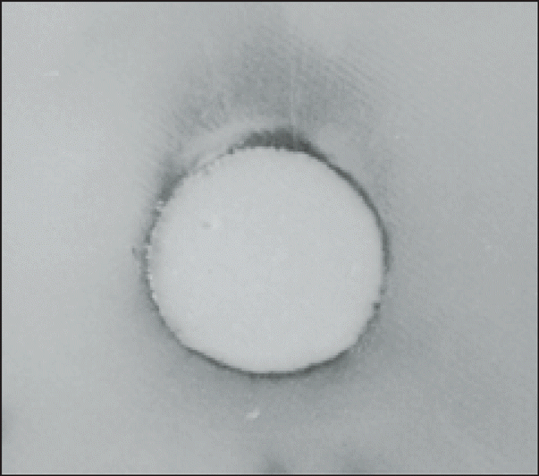
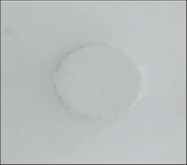
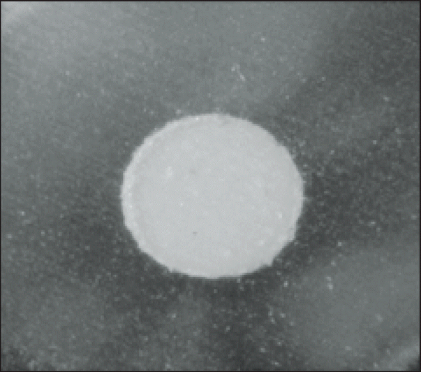
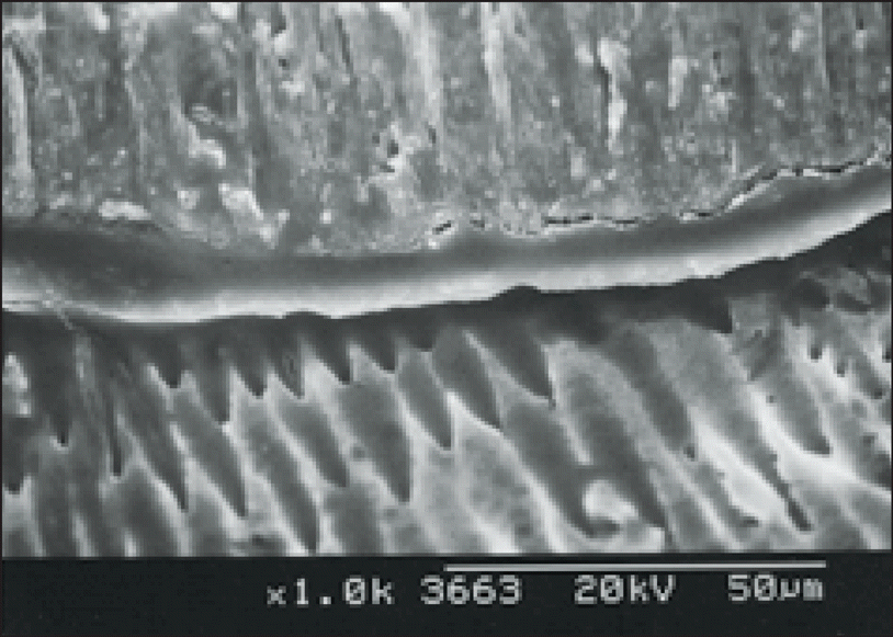 | Figure 18.Photograph of the NaOCl group Level 2 (× 1000); Scanning electron micrographs of tooth sections showing the gap-free region between gutta-percha and the surface of root canals. Surface generally covered with smear layer. |
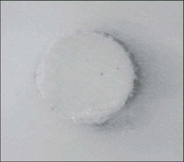
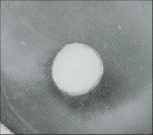
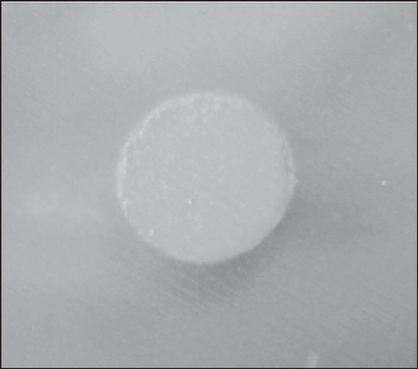
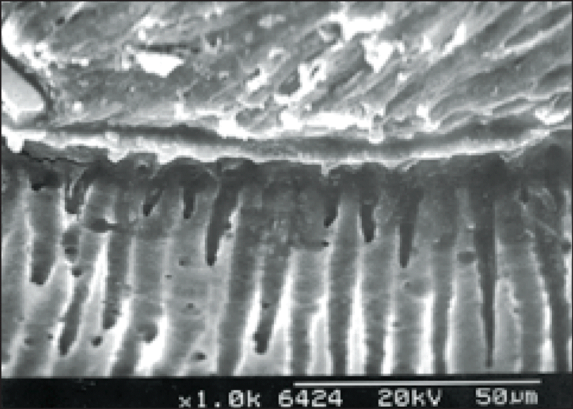
 | Figure 19.Photograph of the EDTA group Level 1 (× 1000); Scanning electron micrographs of tooth sections showing the penetration of sealers to opened dentinal tubules. Gap-free region was showing between the sealer and the dentinal tubules. Numerous sealer tags could be seen within the intertubular dentin. But, voids were occuring between the sealer and the gutta-percha. |
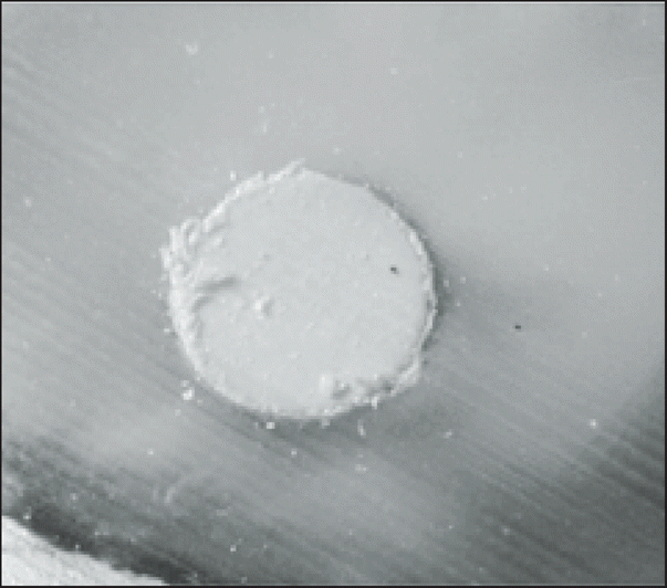
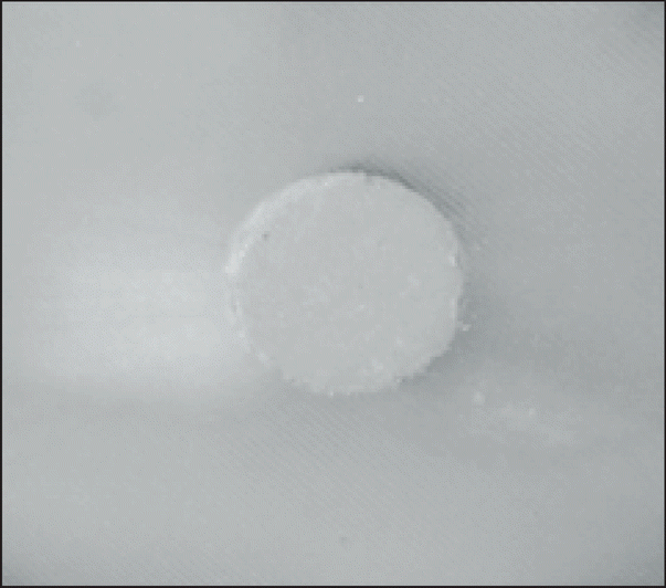
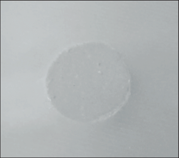
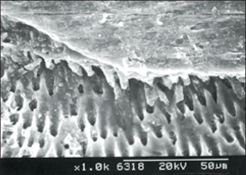
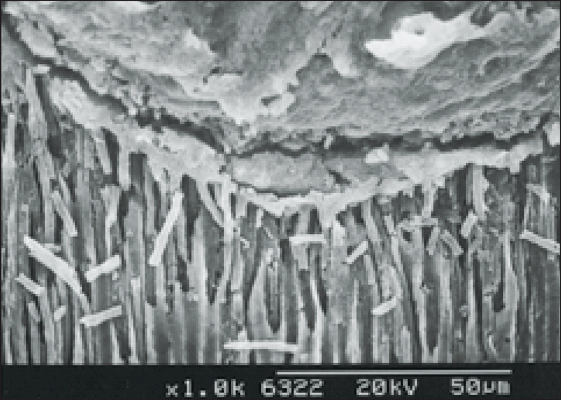 | Figure 20.Photograph of the EDTA group Level 2 (× 1000); Scanning electron micrographs of tooth sections showing the penetration of sealers to opened dentinal tubules. Gap-free region was showing between the sealer and the dentinal tubules. Numerous sealer tags could be seen within the intertubular dentin. |
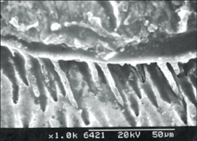
 | Figure 21.Photograph of the NaOCl-6 group Level 1 (× 1000); Scanning electron micrographs of tooth sections showing the gap-free region between gutta-percha and the surface of root canals. Surface generally covered with smear layer. |
 | Figure 22.Photograph of the NaOCl-6 group Level 2 (× 1000); Scanning electron micrographs of tooth sections showing the gap-free region between gutta-percha and the surface of root canals. Surface generally covered with smear layer. |
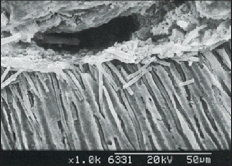
 | Figure 23.Photograph of the EDTA-6 group Level 1 (× 1000); Scanning electron micrographs of tooth sections showing the penetration of sealers to opened dentinal tubules. A few sealer tags could be seen within the intertubular dentin. |
 | Figure 24.Photograph of the EDTA-6 group Level 2 (× 1000); Scanning electron micrographs of tooth sections showing the penetration of sealers to opened dentinal tubules. A few sealer tags could be seen within the intertubular dentin. |
 XML Download