

 PDF
PDF ePub
ePub Citation
 Print
Print


I. INTRODUCTION
Composite resin restoratives have been widely applied in clinical dentistry and replaced amalgam since their introduction1). As a core material, the use of composite is more common nowadays. Composite cores can be classified in three groups by their curing mode. Self-cured type is convenient to build up the core and reduces the chairtime, while it takes a long time to have hardness enough to prepare crown. Light-cured type has advantages to maximize working time and minimize setting time, however, there is a possibility of incomplete polymerization in deep cavities. Recently, dual-cured type that combines the advantages of self-cured and light-cured composites has been introduced and their use has been increased.
Many clinicians used dual-cured type composite core materials in a bulk filling technique. But one of the limitations of these materials is that a hard-top surface is not an indication of adequate polymerization throughout the depth of the restoration in a deep cavity2-6). The effectiveness of polymerization or depth of cure of light-activated composites is vital to ensure optimum physiomechanical properties1,2,4,7). It depends not only the chemistry of the material, but also the filler particle type, size, and quantity1-3). In addition, polymerization is dependent on the effectiveness of the radiation sources, including spectral distribution, intensity, exposure time, and alignment of the light-tip guide1,2,4).
The effectiveness of polymerization may be assessed directly or indirectly1-4,8). Fourier-transformed infrared spectroscopy or Raman spectroscopy is the most commonly used method directly to measure the degree of conversion of resin or composite1,2,7). However, this method is complex, expensive and time consuming1-4,8). Indirect methods have included scraping, visual and surface hardness1-4,9). DeWald and Ferracane compared four commonly used methods for evaluating depth of cure in light-activated composites1-3,10). They found that visual and scraping methods correlated well, but severely overestimated depth of cure as compared with hardness testing or degree of conversion1-3). Hardness testing appears to be the most popular indirect method for investigating factors that influence effectiveness of polymerization because of the relative simplicity of the method and a good correlation between the results of hardness and infrared spectroscopy experiments2,3).
According to a series of recent researches with dual-cured resin cements, enough polymerization occurs at top surface, but did not necessarily occur at bottom surface when light did not reach bottom6). Also, degree of conversion was different according to cure mode in dual-cured resin cements6). In clinical cases of deep cavities, bulk filling with dual-cured resin doesn't show exactly whether polymerization is completed or not in bottom surface.
Therefore the purpose of this study was to investigate the influence of thickness on the degree of cure when bulk filling was done with dual-cured composite core and whether there was enough polymerization occurred at the deep bottom surface by self cure ability.
II. MATERIALS AND METHODS
Two composites core materials, Luxacore Dual and Luxacore Self (DMG Inc, Hamburg, Germany) of shade blue were chosen (Table 1).
The composite restoratives under test were placed in Teflon mold 10 mm in diameter and cylindrical cavities 2, 4, 6, 8 mm in depth4). The material placed in the mold cavity was confined between opposing cellulose acetate strips (KerrHawe striproll, Switzerland). A glass slide was placed on the mold and excess material was extruded out by applying pressure. The materials were irradiated from the top surface through the glass slide as recommended by the manufacturer with a halogen light curing unit (VIP, BISCO Inc, Schaumburg, IL, U. S. A.) at 500 mW/cm2. (Table 2)
In Luxacore dual, the curing time was decided by the thickness of composite, e.g., 20 seconds in 2 mm thick group and 40 seconds in 4 mm group. In 6 mm thick specimens, according to manufacturers' instruction, lower 4 mm incremental layer was cured for 40 seconds then upper 2 mm was added and cured for 20 seconds more (total 60 sec). In 8 mm thick specimens, lower and upper 4 mm layers were cured for 40 seconds each.
In bulk filling groups, 6 mm thick specimens were cured for 60 seconds and 8 mm specimens for 80 seconds at a time for recognizing the effect of self curing in the deep cavity with diminished light. As a control group of self-cure mode of Luxacore Dual, three of each 2, 4, 6, 8 mm specimens were made and cured in a dark room for 30 minutes. In Luxacore Self groups, they were filled with a bulk placement and cured for 4 minutes before removal.
After cured, the specimens were removed from the mold and stored in the dark water bath at 37℃ for 24 hours and the top and bottom surfaces of each specimen were minimally polished with a #2000 sandpaper5). The specimens were positioned centrally beneath the indentor of a digital microhardness tester (Future-Tech Corp, Tokyo, Japan) to assess Knoop's hardness (KHN) of the top and bottom surfaces. A 25 gf load was applied through the indentor with a dwell time of 10 seconds. Ten specimens were made for each material and cavity-depth combination. For each upper and bottom surface, three readings were taken at three points.
Mean KHN at the top and bottom surfaces represented the hardness of each sample and the hardness ratio (KHN of Bottom/Top) was calculated. The statistical analysis was performed using one-way analysis of variance (ANOVA) and Tukey's test at a p = 0.05 significance level.
III. RESULTS
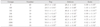
The mean KHN of Luxacore self and dual for the different thickness, curing and filling groups was shown in Tables 3, 4 and 5. For Luxacore self and Luxacore dual-self curing mode groups, there was no significant difference in KHN among the different depths at the top & bottom surfaces (Table 3 and 4), and they are analyzed as one groups (S and DS) in Table 5. S and DS groups showed lower hardness value than the other groups at top surface. At the bottom surface, they had lower hardness value than D2, D4, DI6, and DI8 groups but same with DB6 and higher than DB8.
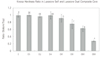
The hardness ratios of the restoratives at the different cavity depths are shown in Figure. DI8, DB6 and DB8 groups showed ratio lower than 0.8, it means polymerization at the bottom surface is not sufficient.
IV. DISCUSSION
Bulk placement of composites may be anticipated in deep cavities and the restorations of endodontically treated teeth if composite resin is used as an amalgam substitute2). It is, however, difficult for visible light to penetrate thick layers of composites, particularly those with darker shades2,8). Filler particles, colorants, and pores scatter visible light and reduce curing efficacy2). The effective intensity of light for curing is dependent on the intensity produced by the curing unit, the distance of the light curing tip from the surface of the material, and internal light scattering within the composite2,3,11). The manufacturers of the Luxacore composites claimed that their products were formulated to be placed in bulk increments of 4 mm with a single 40-second visible-light exposure.
Bulk placement of composites may generate higher internal stresses in the material, more loss of marginal integrity and polymerization shrink-age as the volume of composite to be polymerized became larger.2,12) The degree to which light-activated composites polymerize is proportional to the amount of light to which they are exposed1,2,13). At the upper surface of the composites, where no overlying composites interfere with light transmission, it has been found that even a curing source with relatively low intensity can cure the resin matrix to an extent almost equal to that when the high-intensity lights are used1-3,14).
In the ideal situation, the degree of polymerization of the composite should be the same through-out its depth, and the hardness ratio should be equal to 1 : 1 or very close to it1-4). That is, if polymerization was effective (i. e., maximum cure of the specimen was achieved) for the depth concerned, the ideal ratio should be 1 : 1, as the hardness of the bottom surface should be similar to that of the top surface1,2).
As light passes through the bulk of the composite, the light intensity is greatly reduced due to light scattering, thus decreasing the effectiveness of polymerization1-4,15). This scattering of light accounts for the differences in hardness between the top and bottom surfaces of the composites evaluated when specimens were 2 mm deep1,2,4). Following the previous research for the light curing composites, thickness of composites cured at one time should not be greater than 2 mm to provide uniform and maximum polymerization despite manufacturers'claims2-4).
It has been suggested that the top-to-bottom hardness gradient should not exceed 10 - 20% (ie, hardness ratio should be greater than 0.8) for adequately polymerized photo-activated resin composite1-5,8,16). Depth at which a 20% reduction had taken place in maximum hardness was considered as a realistic depth of cure2,3,17). At depths greater than 2 mm, poor polymerization of composite resulted, for polymerization was very susceptible to changes in light intensity and exposure duration1-3,8,13).
Based on a hardness ratio of 0.8 as a guideline of adequate polymerization of bottom surface, Luxacore self was not affected by the thickness. In Luxacore dual, polymerization was effective in D2, D4 and DI6 specimens but the DI8 group showed just lower than the 0.8 ratio. The results of DB8 were unexpected because the hardness of bottom surface was lower than that of DS group. It could be explained that the initial low intensity light curing accelerated change of the dual cure composite matrix from the gel to postgel phase, thus the free movement of the radical was inhibited18).
In clinical situation, it is very common to meet the cavity deeper than 4 mm. Following the result of the 6 mm groups, incremental filling group showed much higher hardness ratio than bulk filling group. Even in 8 mm depth, if incremental technique is applied, the bottom of the core can be polymerized close to the ratio 0.8. Within the limitation of this experiment, incremental filling is better than bulk filling in case of over 4 mm depth, and bulk filling should be avoided.
When comparing the Luxacore self and dual, the hardness of Luxacore dual-cured composite was greater than that of Luxacore self-cured type. Even though the hardness did not indicate all mechanical properties of the composite, if the both system used the same matrix and filler, light cure groups might have higher physical properties than self cure groups.
V. CONCLUSIONS
In this study, we investigated the influence of thickness on the degree of cure of self- and dual-cured composite core materials when light and self activated.
In self-cure mode, polymerization is not affected by the thickness both in the Luxacore Self and Dual. In light cure mode, however, when the thickness is over 4 mm, incremental filling is recommended to obtain adequate polymerization of the bottom surface.


 XML Download