

 PDF
PDF ePub
ePub Citation
 Print
Print


I. INTRODUCTION
For more than 100 years, gutta percha has been the most commonly used material to obturate the root canal system. It fulfills many of the requirements as a root canal filling material suggested by Grossman1). One of characteristics for the ideal canal filling is its retrievability. Endodontic retreatment is indicated when initial root canal treatment has failed and the problems may be corrected through further canal debridement and obturation. In this aspect, all root canal filling materials should be removed by standardized techniques2).
There have been several studies that investigated the efficient ways of removing gutta percha and sealer using different methods3-10). Techniques described for gutta percha removal included the use of rotary instruments, heat carriers and solvents. In many studies the use of NiTi rotary instruments has been recommended for gutta percha removal and various studies have reported its efficacy, cleaning ability and safety. Hulsmann and Bluhm6) demonstrated that ProTaper rotary instruments were time saving for removing gutta percha. They also showed that the use of solvent was not significantly effective in removing the filling material from the root canal.
Recently, a new root canal filling material was introduced. Resilon (Epiphany, Pentron, Wallingford, CT, USA) is a thermoplastic synthetic polymer-based root canal filling material containing bioactive glass and radiopaque fillers11). According to the manufacturer's claim, it performs like gutta percha and has similar handling properties. Epiphany sealer is a dual curable resin composite sealer with various fillers. Resilon is emerging as an alternative to gutta percha and has been used clinically in many practices for more than 2 years. Regardless of the manufacturer's claims, it is expected that the removal of this material will be necessary in some situations. However, there are only few studies regarding the removal efficacy of this new filling material12-14).
The purpose of the present study was to determine the retrievability of Resilon compared with conventional gutta percha and sealer obturation.
II. MATERIALS AND METHODS
Preparation of the teeth
Twenty-seven extracted human anterior teeth and premolars were obtained and stored in normal saline after sterilization. The total root length was adjusted to 12 mm by removing a part of crown portion. A radiograph was taken for each tooth and only roots with radiographically visible single canals were selected. A size 10 K-file was passed 0.5 mm beyond the apex under the microscope (Carl-Zeiss, Oberkochen, Germany) and the working length recorded as being 1.00 mm less than that length.
All canals were prepared by the same operator using a standardized manner. Sizes 4,3 and 2 Gates-Glidden burs were used for coronal flaring. The canals were instrumented with sizes 15 and 20 K-files to the working length. This was followed by preparation with a series of ProFiles (Dentsply, Tulsa, OK, USA) rotated at 300 rpm. Preparation was completed when a 0.04 taper ProFile with a tip equivalent to ISO size 35 reached the working length. All canals were irrigated with 3.0% NaOCl and 17% EDTA. RC Prep (Premier, Plymouth Meeting, PA, USA) was used as a lubricant.
The teeth were randomly divided into two groups to receive either gutta percha or Resilon as the obturation material.
Group 1 (n = 12): obturation using gutta percha and AH 26 plus sealer
A fine-medium gutta percha cone was trimmed using a gutta gauge (Dentsply, Tulsa, OK, USA) to fit at the working length or at most 1.0 mm short from the working length. An equivalent sized system B plugger (SybronEndo, Orange, CA, USA), prefitted to the 4 mm short of the working length,was selected. Canals were dried with paper points and the gutta percha cone was lightly coated with AH 26 plus sealer (Dentsply, Tulsa, OK, USA). The system B unit was set at 230℃ and power 10 for obturation. After inserting the system B plugger to cut the coronal part of gutta percha, downpacking was performed to the previously determined length. Canals were backfilled with Obtura II (Spartan, Fenton, MI, USA) and condensed with S-Kondensors (Spartan, Fenton, MI, USA).
Group 2 (n = 15): obturation using Resilon
Preparation of the canals before obturation was the same as group 1 except for the final rinse. Instead of 3.0% NaOCl, 2% Chlorhexidine was used as a final irrigant. Obturation was done following the manufacturer's instructions. The self-etching primer (Epiphany Primer) was introduced into the canals with paper points to coat the root canals walls. In 25 seconds, excess primer was removed with new dry paper points. Then, fine-medium sized Epiphany core was applied into the canal after being coated with Epiphany sealer. The system B unit was set at 150℃ and power 10 for obturation. For backfilling Epiphany pellet was inserted to Obtura II unit and the temperature setting was 150℃.
Retreatment
The samples were kept at 37℃ and 100% humidity for 7 days after the coronal and apical portion was sealed with utility wax.
Initially, sizes 4,3 and 2 Gates-Glidden burs were used to remove the coronal portion of the filling material. ProFiles were run at 500 rpm to remove the remainder. Light apical pressure was applied to work the files apically to the working length. RC Prep and 3% NaOCl were used during the instrumentation. Canals were enlarged to one size larger than the previous master apical size. The total time for retreatment was recorded commencing after the initial removal of filling material with Gates Glidden burs and ending when canals were instrumented by an ISO size 40, 0.04 taper ProFile.
After final instrumentation, all canals were irrigated with 3.0 ml of 3% NaOCl, soaked with 1.0 ml of 17% EDTA for 5 minutes and finally rinsed with 3.0 ml of sterile water. The time required for the final irrigation was not included in the total retreatment time.
Sample analysis using SEM
The teeth were grooved vertically with burs and discs in the buccal and lingual surfaces. After being split longitudinally with a chisel, the samples were prepared for scanning electron microscopy. General cleanliness (× 75) of the coronal, middle, and apical thirds was evaluated using 5 scoring system:
1 : clean, less than 10% of surface was covered by debris
2 : 10 - 30% of surface was covered by debris
3 : 30 - 60% of surface was covered by debris
4 : 60 - 90% of surface was covered by debris
5 : more than 90% of surface was covered by debris
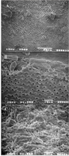
For selected representative samples from each group, the observation was performed with higher magnifications (× 500 - 1,500) to examine whether the dentinal tubules were patent after filling materials were removed.
Statistical analysis
Time required for material removal in two groups was measured in minutes and expressed as mean ± SD. Group comparison was done using a Student t-test. A Chi-square analysis was performed to analyzed canal cleanliness and debris removal. A p value of < 0.05 was used to determine significance. All sample preparation, treatment was performed by a single operator. Evaluation for cleanliness was done by two dental students after calibration, and they made an agreement on each SEM picture.
III. RESULTS
Time required for complete removal of filling material
Time for retreatment in group 1 which was filled with gutta percha and AH 26 plus sealer was 3.25 ± 0.32 minutes. In group 2, removing the Epiphany core and sealer from the canal took 3.05 ± 0.34 minutes. There was no statistically significant difference between two experimental groups (p > 0.05).
Cleanliness of root canal walls
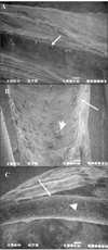
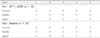
The results for root canal cleanliness are summarized in Table 1. There was no significant difference between the two experimental groups. Generally most of the specimens demonstrated clean surface with small amount of sealer. More debris remained in the apical and middle thirds than in the coronal part. The openings of dentinal tubules were detected under higher magnifications in both experimental groups (Figure 2). However, dentinal tubules were not always patent. More smear layer was observed in the apical root wall (Figure 1).
IV. DISCUSSION
Removing as much sealer and filling material as possible may be critical for the success of retreatment. In this study for removing the previous obturation, Gates-Glidden burs and ProFiles were used. There have been several studies evaluating the usage of NiTi rotary files in conventional retreatment3-10). Even though Barrieshi-Nusair4) showed that the use of SS hand file was faster compared with NiTi files in gutta percha removal, most researchers reported that NiTi rotary files were efficient to retrieve old canal filling materials3,5).
Speed set up for removing gutta percha was slightly variable depending on the instrument types and operators. For example, Bramante and Betti15) used Quantec rotary files at 1500 rpm for filling material removal. On the other hand, Ferreira et al.5) used ProFiles rotated at 300 rpm for gutta percha removal. In the present study speed was adjusted to 500 rpm for removing the filling materials.
After material removal, more debris remained in the apical and middle thirds than in the coronal part. This is in accordance with other studies on gutta percha removal techniques. Masiero and Barletta8) reported the apical third had the most remaining material regardless of removal technique. Also Kosti et al.7) claimed that none of the methods used for the removal of root filling was totally effective, especially in the apical third. The result for this study demonstrated that canal enlargement to one size larger than original preparation might not be enough to render the dentinal wall clean and the tubules patent.
Ezzie et al.13) showed that Resilon was faster to remove than gutta percha. And de Oliveira et al.12) reported that the mean time required for removing gutta percha/AH 26 sealer and Resilon was 1.10[SSJ1] minutes and 0.89 minutes respectively. However, no significant difference was found in the efficacy of retreatment between gutta percha and Resilon groups in the present study.
As a summary the study showed that Resilon was effectively removed by Gates Glidden burs and ProFiles. Its general handling properties for retrieval were similar to those of gutta percha.
 XML Download