

 PDF
PDF ePub
ePub Citation
 Print
Print


I. Introduction
The objective of root canal preparation is to clean and shape the root canal system, while maintaining the original configuration. A continuously tapering, conical, funnel-shaped canal with the smallest diameter at the end point and the largest at the orifice has been perceived to be the most appropriate for filling with gutta-percha1).
In small curved canal, it is difficult to achieve this object. During root canal preparation, aberrations such as apical zipping, canal transportation, ledge, and strip perforation often occur, which inversely influence on the treatment success1). Tendency of files to straighten in the canal removes excessive amount of dentin from the inner wall of the curvature at the curve and the outer wall of it at the root end2,3). This effect causes straightening the canal and thus may shorten the working length.
Many techniques and various instruments have been developed in an attempt to improve the quality of root canal treatment in curved canals. Anticurvature filing4), step-back filing2), crown-down pressureless filing5), and balance forced technique6) were introduced. In spite of improvements in instrument design and in various root canal preparation methods, stainless steel hand files still tend to create a number of aberrations in curved canals2,7).
During the last decade, various kinds of root canal instruments made of nickel-titanium alloys were developed. Nickel-titanium instruments were found to have 2 to 3 times more elastic flexibility of the stainless steel files in bending and torsion, as well as superior resistance to torsional fracture8). Nickel-titanium files were more effective in maintaining the original canal path of curved root canal7,9-11). Different kinds of canal instrument and canal preparation technique may exert different effects on working length. If working length is shortened during the course of treatment, overinstrumentation and overfilling may occur. This may cause error of canal shaping and delay healing of periapical lesion.
Therefore, the aim of this study was to evaluate the change of working length with various instrumentation techniques in curved canals.
II. Materials and Methods
Buccal canals of maxillary molars and mesial canals of mandibular molars were used. Using the technique described by Schneider12), canal curvature of root was determined and recorded. Canals having curvature above 20 degrees were selected.
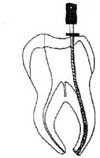
Occlusal surface of teeth was flattened by using diamond point and sandpaper. After access cavity preparation, lingual surface of teeth were embeded in acrylic resin so that the maximum of canal curvature can be seen and root apex was flattened to facilitate reproducible accurate measurements (Figure 1).
Forty specimens were divided into four groups according to the two kinds of hand instruments and two kinds of rotary instruments used: stainless steel hand file (MANI®, Matsutani Seisakusho Co., Takanezawa, Japan), nickel titanium hand file (Naviflex NT™, Brassler, Savannah, USA), ProFile®, and ProTaper™ (Densply-Maillefer, Ballaigues, Switzerland). A #10 K-file was introduced into the canal of each specimen and was placed against a flattened root apex using stereomicroscope. Rubber stop was fixed with a glue and working length was determined. Each canal was instrumented until apical diameter had attained a size of 30. Root canals were irrigated with NaOCl and RC-Prep (Stone Pharmaceuticals, Philadelphia, USA) was used as a lubricant.
In two hand file groups, #2 and #3 Gates Glidden drill were used to flare the coronal third of the canal and crown-down pressureless technique5) was used. A #30 file was placed into the canal until resistance was first encountered and whole procedure was used until apical file size of #30 was obtained.
In ProFile group, Orifice Shaper #3 and #2 were used until resistance was encountered (12 to 15 mm) for coronal flaring. The .06/#25 and .06/#20 files were used serially to the resistance (within 1 to 2 mm of the working length). For apical shaping, .04/#25 and .04/#30 files were used to the working length.
In ProTaper group, SX, S1, and S2 files were used for coronal flaring. S1 was used first and moved apically to 2 mm short of the working length. SX files were then used sequentially until resistance was encountered (4 to 5 mm from the working length), followed by S1 and S2 to the working length for the shaping of the coronal two thirds of the canal. The apical one third was finished by using F1, F2 and F3 sequentially to the working length, with only one pecking motion for each instrument.
Working length change was examined before and after finishing the coronal flaring and after finishing the apical preparation. Positional relation between the tooth apex and the file tip was examined before and after coronal flaring and after apical preparation under a stereomicroscope, and the images were stored in a computer using a CCD camera and micro VIDEO Studio 200 program. The change of file tip position was measured by using AutoCAD 2000 (Autodesk Corp., San Rafael. CA, USA).
Root canal curvature was measured in radiograph with K-file in the canal using Schneider's method12) before and after coronal flaring, and after apical preparation.
The data were analysed using one-way ANOVA and Tukey's studentized range test.
III. Results
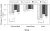
Working lengths were decreased significantly in each stage in all instrumentation groups. Coronal flaring using Gates Glidden burs in hand file groups caused significantly more working length change than in ProFile instrumentation group (p < 0.05) (Figure 2).
Whole canal instrumentation in stainless steel hand file group caused significantly more working length decrease than in ProFile instrumentation group (p < 0.05) (Figure 2).
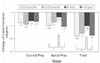
Canal curvature was decreased in all groups. There was no significant different in the change of canal curvature after coronal flaring among groups.
Apical preparation using either stainless steel hand file or ProTaper caused significantly more decrease of canal curvature than in ProFile (p < 0.05).
Whole canal instrumentation in either stainless steel hand file group or ProTaper group caused significantly more decrease of canal curvature than in ProFile instrumentation group (p < 0.05) (Figure 3).
IV. Discussion
In the present study, working length was decreased in all canals, indicating that shortening of working length occurs during the instrumentation in curved canals. A number of studies reported shortening of working length13-17). Schroeder et al.16) reported that change of working length by Gates Glidden drill was 0.17 mm. In the present study, change of working length by coronal flaring is similar with the result of Schroeder et al.16). The finding that ProFiles caused the smallest change in working length are in agreement with those of Thompsom and Dummer18,19), Bryant et al.20,21), and Kum et al.22) who evaluated length change associated with the use of nickel-titanium rotary files. According to Thompson and Dummer18,19), Bryant et al.20,21), and Kum et al.22), nickel-titanium rotary files caused minimal change in canal configuration.
In the present study, change of working length and change of canal curvature were measured. Coronal flaring using Gates Glidden burs in hand instrument groups and whole canal instrumentation in stainless steel hand file group caused significantly more working length change than in ProFile instrumentation group. This result is also similar with the report of Davis et al.17). Nickel-titanium files were more effective in maintaining the original canal path of curved root canals, so this is reasonable result. Also, change of canal curvature in ProFile group after apical preparation and whole canal instrumentation significantly smaller than in stainless steel hand file group and ProTaper group.
Kim et al.15) reported that decrease of working length and canal curvature occurred during canal preparation using ProFile and greatest change of curvature and working length were observed in above 30 degrees.
In the present study, working length and curvature was decreased in all groups. Significantly smaller decrease in curvature was observed in ProFile group than in stainless steel K-file group and ProTaper group. Since the group of the greatest change of canal curvature showed the greatest change of working length as well, and the group of the smallest change of canal curvature showed the smallest change of working length as well in the present study, the main cause of the change of working length will be the change of canal curvature. Because of decrease of curvature, working length may be decreased, and also file didn't passed by center of canal and was straightened in canal, this may cause decrease of working length. Therefore, decrease of working length will occur even when decrease of curvature don't observed. In the present study, because canal curvature was determined by measuring curvature of file, change of canal curvature in this study may be different from the exact change of canal curvature and may be greater than that.
In response to the problems with the traditional step-back preparation method, there has been a trend toward the crown-down technique, which involves preparing a canal from the cervical aspect to the apex, rather than the classical approach from the apex to the crown23).
The crown-down technique using nickel-titanium files with increased tapers allows easier access to the canal, improves distribution of irrigant and induces less extrusion of debris than other technique11).
ProTaper is a Ni-Ti rotary file with high taper and high cutting ability. ProTaper removed significantly more canal material than ProFile, GT file, and Quantec, and cause decrease in canal curvature. ProTaper removed more material from the inner side of canal before the initiation of curve, but in the apical third area, they removed more material from the outer side of canal, which resulted in a decrease in curvature24). ProTaper removed more root dentin than GT Rotary, Quantec, and ProFile at danger zone25). Therefore, canal preparation using ProTaper may cause greater shortening of working length than other Ni-Ti instruments.
If working length is shortened during the course of treatment, overinstrumentation and overfilling may occur. This may cause error of canal shaping and delay healing of periapical lesion. In the present study, both the coronal flaring and the apical preparation caused change of working length in each group, and change of working length by coronal flaring was greater than that by apical preparation. Therebefore, using the working length measured before root canal preparation may cause overinstrumentation. Hence, if an operator wants to measure working length only once, it will be recommended to do it after coronal flaring rather than before root canal preparation.
The result of the present study demonstrates that working length and canal curvature decrease with all kinds of instrumentation techniques. Therefore, special caution is needed to minimize them and redetermination of working length will be necessary before final instrumentation. Further research is needed to evaluate the change of working length and apical canal shape with various designs of endodontic instruments.
 XML Download