Abstract
This study was conducted to evaluate canal configuration after shaping by ProTaper™ with various rotational speed in J-shaped simulated resin canals.
Forty simulated root canals were divided into 4 groups, and instrumented using by ProTaper™ at the rotational speed of 250, 300, 350 and 400 rpm. Pre-instrumented and post-instrumented images were taken by a scanner and those were superimposed. Outer canal width, inner canal width, total canal width, and amount of transportation from original axis were measured at 1, 2, 3, 4, 5, 6, 7 and 8 mm from apex. Instrumentation time, instrument deformation and fracture were recorded. Data were analyzed by means of one-way ANOVA followed by Scheffe's test.
The results were as follows
Regardless of rotational speed, at the 1~2 mm from the apex, axis of canal was transported to outer side of a curvature, and at 3~6 mm from the apex, to inner side of a curvature. Amounts of transportation from original axis were not significantly different among experimental groups except at 5 and 6 mm from the apex.
Instrumentation time of 350 and 400 rpm was significantly less than that of 250 and 300 rpm (p < 0.01).
In conclusion, the rotational speed of ProTaper™ files in the range of 250~400 rpm does not affect the change of canal configuration, and high rotational speed reduces the instrumentation time. However, appearance of separation and distortion of Ni-Ti rotary files can occur in high rotational speed.
Figures and Tables
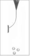
 | Figure 1Pre-instrumentation image. Working length was 16 mm, the round circles were the landmark for getting the superimposed image. |
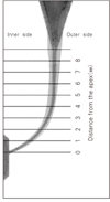
 | Figure 2The diagram indicates the points at which the canal width was measured after superimposition of pre-instrumentation and post-instrumentation image. |
References
1. Weine FS, Kelly RF, Lio PJ. The effect of preparation procedure on original canal shape and on apical foramen shape. J Endod. 1975. 1:255–262.

2. Schilder H. Cleaning and shaping the root canal. Dent Clin North Am. 1974. 18:269–296.
3. Skidmore AE, Bjorndal AM. Root canal morphology of the human mandibular first molar. Oral Surg Oral Med Oral Pathol. 1971. 32:778–784.

4. Abou-Rass M, Frank AL, Glick DH. The anticurvature filing method to prepare the curved root canal. J Am Dent Assoc. 1980. 101:792–794.

5. Bramante CM, Berbert A, Borges RP. A methodology for evaluation of root canal instrument. J Endod. 1987. 13:243–245.
6. Walia H, Brantley WA, Gerstein H. An initial investigation of the bending and torsional properties of nitinol root canal files. J Endod. 1988. 14:346–351.

7. Stoeckel D, Yu W. Superelastic Ni-Ti wire. Wire J Int. 1991. 3:45–50.
8. Glossen CR, Haller RH, Dove SB, del Rio CE. A comparison of root canal preparations using Ni-Ti hand, Ni-Ti engine-driven, and K-Flex endodontic instruments. J Endod. 1995. 21:146–151.

9. Thompson SA, Dummer PMH. Shaping ability of Hero 642 rotary nickel-titanium instruments in simulated root canals: Part 1. Int Endod J. 2000. 33:248–254.

10. Park HS, Lee MK, Kim JJ, Lim YJ, Jang MS, Lee JY. A study on the shape of a canal prepared with 'three-file' technique in a curved canal. J Korean Acad Conserv Dent. 2000. 25:494–497.
11. Coleman CL, Svec TA. Analysis of Ni-Ti versus stainless steel instrumentation in resin simulated canals. J Endod. 1997. 23:232–235.

12. Kim YT, Baek SH, Bae KS, Lim SS, Yoon SH. A comparison of three stainless steel instruments in the preparation of curved root canals in vitro. J Korean Acad Conserv Dent. 2001. 26:9–15.
13. Esposito PT, Cunningham CJ. A comparison of canal preparation with Nickel-Titanium and Stainless steel instruments. J Endod. 1995. 21:173–176.

14. Bolanos OR, Jensen JR. Scanning electron microscope comparison of the efficacy of various methods of root canal prparation. J Endod. 1980. 6:815–821.
15. Calhoun G, Mongomery S. The effects of four instrumentation technique on root canal shape. J Endod. 1988. 14:273–277.
16. Dummer PMH, Alodeh MHA. A method for the construction of simulated root canals in clear resin blocks. Int Endod J. 1991. 24:63–66.

17. Kum KY, Spangberg L, Cha BY, Jung IY, Lee SJ, Lee CY. Shaping ability of three ProFile rotary instrumentation techniques in simulated resin root canals. J Endod. 2000. 26:719–723.

18. Peters OA, Peter CI, Schonenberger K, Barbaknow F. ProTaper rotary root canal preparation: effects of canal anatomy on final shape analysed by micro CT. Int Endod J. 2003. 36:86–92.

19. Veltri M, Mollo A, Pini PP, Ghelli LF, Balleri P. In vitro comparison of shaping abilities of ProTaper and GT rotary files. J Endod. 2004. 30:163–166.

20. Yun HH, Kim SK. A comparison of the shaping abilities of 4 nickel-titanium rotary instruments in simulated root canals. Oral Surg Oral Med Oral Pathol Oral Radiol Endod. 2003. 95:228–233.

21. Koch K, Brave D. The ultimate rotary file? Oral Health March. 2002. 59–64.
22. Iqbal MK, Firic S, Tulcan J, Karabucak B, Kim S. Comparison of apical transportation between ProFile and ProTaper NiTi rotary instruments. Int Endod J. 2004. 37:359–364.

23. Karagoz-Kucukay I, Ersev H, Engin-Akkoca E, Kucukay S, Gursoy T. Effect of rotational speed on root canal preparation with Hero 642 rotary Ni-Ti instruments. J Endod. 2003. 29:447–449.

24. Poulsen WB, Dove SB, Del Rio CE. Effect of nickel-titanium engine-driven instrument rotational speed on root canal morphology. J Endod. 1995. 21:609–612.

25. Calberson FL, Deroose CA, Hommez GM, De Moor RJ. Shaping ability of ProTaper™ nickel-titanium files in simulated resin root canals. Int Endod J. 2004. 37:613–623.


 PDF
PDF ePub
ePub Citation
 Print
Print


 XML Download