

 PDF
PDF ePub
ePub Citation
 Print
Print


Abstract
During root canal preparation procedures, the potential for instrument separation is always present. Files, a lentulo, a Gates-Glidden (GG) bur or any manufactured obstruction can be left behind in the canal. Nickel-titanium (NiTi) rotary files are in common usage in these days. Despite their undeniable advantages, there is a potential risk of separation within the canals. It is very rapid, unpredictable, and creates a great deal of stress for the practitioner.
When an endodontic instrument separates, the best option is to remove it. Ultrasonic instruments and microscopes have improved the success rate for removing separated instruments. But it is difficult and not always possible. Therefore prevention is the key.
In this case report, several management methods of separated file in the canal are presented.
Figures and Tables
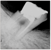
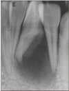
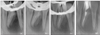
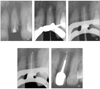
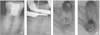 | Figure 1Diagnostic intraoral radiograph (a). Separated file in the mesial root isthmus (b, c) and remove it using ultrasonic device under microscope (d).
|
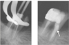
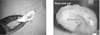
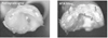
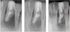
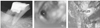 | Figure 3File separation in the root canal (a, b) and remove using ultrasonic device under microscope (c).
|
References
1. Mounce R. The heartache of separation. Dent Today. 2003. 22(4):83–85.
2. Ruddle C. Cleaning and shaping the root canal system. Pathways of the Pulp. 2002. 8th ed. St. Louis: Mosby;231–292.
3. Nagai O, Tani N, Kayaba Y, Kodama S, Osada T. Ultrasonic removal of broken instruments in root canals. Int Endod J. 1986. 19(6):298–304.

4. Hulsmann M, Schinkel I. Influence of several factors on the success or failure of removal of fractured instruments from the root canal. Endod Dent Traumatol. 1999. 15(6):252–258.
5. Ruddle CJ. Broken instrument removal. The endodontic challenge. Dent Today. 2002. 21(7):70–81.
6. Shen Y, Peng B, Cheung GS. Factors associated with the removal of fractured NiTi instruments from root canal systems. Oral Surg Oral Med Oral Pathol Oral Radiol Endod. 2004. 98(5):605–610.
7. Ward JR, Parashos P, Messer HH. Evaluation of an ultrasonic technique to remove fractured rotary nickel-titanium endodontic instruments from root canals: an experimental study. J Endod. 2003. 29(11):756–763.
8. Ward JR, Parashos P, Messer HH. Evaluation of an ultrasonic technique to remove fractured rotary nickel-titanium endodontic instruments from root canals: clinical cases. J Endod. 2003. 29(11):764–767.
 XML Download