

 PDF
PDF ePub
ePub Citation
 Print
Print


I. INTRODUCTION
Root canal filling is an important procedure in endodontic treatment to prevent root canal reinfection for successful endodontic prognosis. Care must be taken during this procedure to avoid root canal cross-infection by instruments or filling materials. Gutta-percha (GP) cones, now widely used to fill root canals, may become contaminated by pathogens during manufacturing and exposed to the air in clinics for several months. It is controversial whether the sterilization process is necessary because of anti-bacterial characteristic of a component of cone itself1,2), however, the sterilization of cones prior to root canal filling has been recommended in most cases.
The chemical agents that have been used to sterilize GP cone prior to canal filling are diverse. Recently, numerous experiments have been performed to seek chemical agents that act in a shorter time and sterilize various bacteria, and it has been reported that NaOCl, glutaraldehyde, chlorhexidine, etc. are effective3-7). However, in these experiments, GP cone was infected with artificially selective bacteria and the sterilization effect among different sterilizers was compared. Therefore, it is necessary to identify the microbial species on GP cones exposed at the outpatient clinics and evaluate which chemical solution is more effective on such bacteria.
On the other hand, the physical changes of GP cones after sterilization have been pointed out. Moller et al. have claimed when the cone was treated with 70% isopropyl alcohol, 5% chloramine, and 0.5% chlorhexidine, the tensile strength of the cone decreased evidently, and thus the physical property of gutta-percha cone could be altered by the surface sterilizer8). In addition, Lee et al. reported that from 1 day after the treatment with 70% isopropyl alcohol, 2.5%, and 5% NaOCl, the tensile strength and the elongation rate of the cone (evidently) decreased, and such alteration could mediate an effect during canal filling in clinics9). Therefore, the purpose of this study was first, to identify the type of bacteria that contaminate GP cone exposed at two hospital-based dental clinics by polymerase chain reaction (PCR), and secondly to evaluate the short-term sterilization effect of three chemical disinfectants. It also analyzed the alteration of the surface texture and physical properties after short-term sterilization, particularly the tensile strength and the elongation rate.
II. MATERIALS AND METHODS
1. The measurement of the contamination rate of gutta-percha cone and the identification of bacteria by PCR
150 GP cones (Meta Biomed Co. Chung-Ju, Korea) kept exposed in two hospital-based dental clinics were collected and used in this study. From 10 immediately opened GP packs, total 30 cones were collected and contamination was assessed as control group. Each GP cone was placed in tube containing 200 µl PBS buffer solution, and vortexed for 5 minutes. All GP cones were removed from the tube, 200 µl PBS buffer solution was inoculated to a Brain Heart Infusion (BHI) agar plate, and cultured for 48 hours in a 37℃ incubator. The number of colonies formed from each GP cone (CFU) was counted and the contamination level of total gutta-percha cones was evaluated. The colony pattern formed on the BHI agar plate was examined and classified by microscope.
Polymerase chain reaction (PCR) amplication was performed in a thermal cycler (Perkin-Elmer Inc. Boston, USA) using colony selected from BHI agar plate as a template. Primer, dNTP (Bioneer, Daejeon, Korea), Taq-polymerase (Bioneer, Daejeon, Korea), PCR buffer, and distilled water were added to a PCR tube together. At that time, the universal 16S rRNA primer (TpU1: 5'-AGAGTTTGATCMTGGCTCAG-3', RTU3: 5'-GWATTACCGCGGCKGCTG-3', Bioneer, Daejeon, Korea) was used as a primer for PCR amplication. The PCR conditions used in this study were as follows: the initial denaturation was at 95℃ for 5 minutes. Thirty amplication cycles were then performed: the denaturation reaction at 95℃ for 1 minute, the annealing at 56℃ for 1 minute, and the extension reaction at 72℃. After the electrophoresis and confirming of PCR products, they were purified using PCR purification system (Bioneer, Daejeon, Korea). Automatic nucleic acid sequencer analyzed the purified DNA. Using the nucleic acid sequence of bacteria thus obtained, the name of bacteria was identified using the BLAST program of nucleotide database (NCBI).
2. Evaluation of sterilization effect of three chemical disinfectants
The bacteria identified above were inoculated to BHI liquid medium and cultured at 37℃ for 24 hours. A GP cone sterilized with EO gas was transferred to the BHI medium that bacteria were cultured, and remained in contact with the bacteria for 2 hours to promote surface contamination. The contaminated GP cone was transferred to a petri dish matted with two layers of filter paper and dried for 24 hours at room temperature. A contaminated GP cone was immersed in each chemical solution for 1 minute, 5 minutes, 10 minutes, and 30 minutes, and dried. As chemical disinfectants, 5.25% NaOCl, 2% chlorhexidine, and ChloraPrep one-step were used. ChloraPrep one-step is a mixed solution (vol: 1 : 1) of 75% isopropyl alcohol and 2% chlorhexidine and is a fast acting, broad spectrum, persistent antiseptic that significantly reduces the number of microorganisms on intact skin. After each decontamination method, all the GP cones were transferred to sterile trial tube containing sterile BHI and incubated at 37℃ for 7 days. Growth, as indicated by the turbidity and the subculture of the examined BHI liquid medium, was then recorded. The subculture was performed with the following method: the observed liquid medium 150µl was inoculated to a Brain Heart Infusion (BHI) agar plate, and cultured for 24 hours in a 37℃ incubator. The sterilization was confirmed by colony forming state. The positive control group was the GP cone immersed in the BHI liquid medium that bacteria were cultured, and dried without sterilization, and cultured 24 hours in fresh BHI culture medium. The negative control group was a GP cone sterilized with EO gas without bacterial contamination and cultured in BHI liquid medium as is.
3. SEM observation of the surface texture of GP cone after 5 min soaking in chemical disinfectants
GP cones were immersed in 3 types of chemical disinfectants (5.25% NaOCl, 2% chlorhexidine, ChloraPrep one-step) for 5 minutes. Their surfaces were compared with that of a fresh GP cone by a scanning electron microscope (×500 and ×10000, JSEM-820, Tokyo, Japan).
4. Assessment of the alteration of the physical property of GP cone after chemical sterilization
By using a Universal testing machine (Instron 5500, Canton, USA), the tensile strength and elongation rate of GP cones soaked in 3 types of chemical disinfectants for 5 minutes were measured. In each experiment group, 40 cones were used. During the measurement of the tensile strength and the elongation rate, a rubber coating grip was used, the crosshead speed was 1.5 mm/min, and the room temperature was maintained at 25℃. The statistical association between the type of chemical disinfectants and the change of physical properties of GP cones was determined by One-way ANOVA and Duncan grouping with a significance level of 0.05.
III. RESULT
PCR identification of bacterial species on the surface GP cones
The immediately opened control GP cones showed negative cultures in all cases. Among total 150 GP cones opened in clinics, bacteria were detected on the surface of 29 gutta-percha cones (19.4%). The numbers of GP cone showing total 1-10 bacteria colonies were 13 (8.7%), the cones showing 11-100 bacterial colonies were 12 (8%), and the cones detected over 101 colonies were 4 (2.7%).
In addition, examining the pattern or color of the colonies formed on BHI agar plate, 1 or 2 types of bacteria were detected on each GP cone. From these colonies, 35 PCR products were purified, and finally, 32 bacteria were identified. Most of them were Staphylococcus spp. In particular, Staphylococcus epidermidis (n = 20/32, 62.5%) was the most prevalent one. In addition, Staphylococcus caprae (n = 8/32, 25%), Staphylococcus capitis (n = 4/32, 12.5%), and Staphylococcus xylosus (n = 1/32, 3.1%) were also detected.
The sterilization effect of chemical solutions
Three chemical disinfectants showed sterilization effect after only 1 minute immersion, which was confirmed not only by the measurement of the turbidity but also by the subculture of the liquid culture medium. Detailed results are shown in Table 1.
SEM Observation of the surface texture of GP cone after 5-min soaking in chemical disinfectants
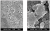
In comparison with GP cone that was not sterilized, the surface of GP cone immersed for 5 minutes in 2% chlorhexidine and ChlorapPrep one-step was slightly wrinkled with the pattern of shrinkage along the long axis of cone, and the adhesion or release of other materials on the surface was not detected. However, on GP cone immersed in 5.25% NaOCl, the precipitate with cuboidal crystal was attached to the overall surface, and it was found to be particularly abundant in the defect area or folded area of cone (Figure 1a). Under ×10000 magnification, a large quantity of round shaped structure was detected in the deep level of defect area of GP cone. It seemed that it was detected as the pattern of the partial lysis of some components of GP cone (Figure 1b).
The alteration of the physical property of GP cone after 5-min soaking in chemical disinfectants
Table 2 & 3 show the average values of the elongation rate and the tensile strength in each experimental group. In regard to tensile strength, the NaOCl-soaked group showed a significant difference than the other group (Table 2, p < 0.05). In terms of elongation rate, all chemical solutions showed significant difference than the control group, especially in the ChloraPrep group (Table 3, p < 0.05).
IV. DISCUSSION
Molecular genetic methods, particularly the polymerase chain reaction (PCR), have been widely used for microbial identification purposes. PCR assays are very sensitive and enable a reliable identification of microbial species or strains that are difficult or even impossible to culture10). In this study, the method of assay should detect the smallest possible number of microorganism. The high sensitivity of the PCR method makes it ideal for this purpose.
Our present study clearly showed that GP cones exposed in clinics can be contaminated by specific organisms, particularly Staphylococcus spp. Staphylococcus epidermidis detected most frequently in this study, is a normal flora residing in the skin and the mucosa of respiratory system in normal individuals. Nevertheless, in immune suppressed patients or in the case at a site different from the distribution of normal flora such as implanted medical devices, or when its number is increased suddenly by other reasons, it may cause severe infection as opportunistic microorganism11).
In a previous study12), GP cones in over-filled areas were covered with a biofilm structure and a colony of cocci was observed in the cracks of the biofilm structure. These findings suggested that cocci were located in deeper layers of the biofilm structure and these cocci might play an important role in initiating biofilm formation. In addition, the biofilm which form on the extra-radicular area of gutta-percha cone are related to refractory periapical pathosis, and gutta-percha cones might play a role in the initiation of biofilm infection in cases of excessive root filling with periapical lesions12,13). Therefore, considering the importance of the aseptic root canal and the contamination of the GP cone in clinics, it may be necessary to decontaminate the gutta-percha cones by means of a chemical disinfectant before canal filling. The time needed for disinfection of GP cone is various according to the types and concentrations of chemical disinfectants or the selective microbial species. It has been reported previously that 1 minute short sterilization with 5.25% NaOCl eradicated spores as well as bacteria. Because of its superior antibacterial effect, the sterilization of the GP cone with 5.25% NaOCl prior to canal filling has been recommended for a long time4-7). However, Short et al. have shown that the cluster of cuboidal crystals on the surface of NaOCl-soaked GP cone was detected at various levels and mentioned that at the time of canal filling, it might affect the apical sealing14). Our present study demonstrated not only cuboidal crystals but also round shaped structures that appeared to be the components of cone on the surface of NaOCl-soaked GP cone under high magnification. Therefore, it is essential to find other chemical disinfectants, which are more stable and clinically useful, to sterilize the contaminated GP cone.
ChloraPrep one-step (2% chlorhexidine gluconate + 70% isopropyl alcohol) is an approved antiseptic for preoperative skin preparation and has the fast bactericidal effect of alcohol and the substantive effect of chlorhexidine. Our results showed that all three chemical disinfectants including ChloraPrep one-step were efficient for sterilization of GP cones by immersion only for 1 minute, which was confirmed by the turbidity measurement and subcultures. We contribute this result to the contamination of GP cone by Staphylococcus, which is a different species from the spore-forming bacteria, fungus with a thick lipid layer, and small virus lacking the envelope that need more powerful sterilization agents. Therefore, GP cone could be easily sterilized in three chemical disinfectants in the present study. Time is more critical when it is necessary to disinfect extra accessory cones during compaction techniques for multi-rooted tooth, because it needs 5 to 10 min for cone disinfection. Therefore, in this study we evaluated the changes of surface textures and physical properties of gutta-percha cones only after 5-min soaking in chemical solutions.
As the factors that alter the physical property of GP cone, the content of gutta-percha, storage conditions, the surface treatment with chemical solutions, etc. have been suggested. Similarly, in the result of our study, it was found that the treatment with chemical solutions affected the tensile strength and elongation rate of gutta-percha cone. Particularly, the result of the treatment with NaOCl was different from that of the study reported by Lee9). This difference is supposed to result from sterilization time. In the study performed by Lee, the period immersed in NaOCl was from 1 day to 30 days and this long-term sterilization treatment generated difference from our results. On the other hand, Valois has shown by atomic force microscopy that only 1 minute treatment with 5.25% NaOCl changed the elasticity of GP cone15).
On root canal filling, the alteration of physical property of GP cone may affect the outcome of root canal treatment. In other words, in the cases of the cone with a high elongation rate, it was thought that the change of the shape of cone itself to irregular canal shape may occur more readily by the same load and it could compact the space between cones more densely. Nonetheless, other physical properties of cone such as modulus of elasticity, compressive strength, etc. have to be considered together. Hence, it is considered that additional clinical studies have to be performed in regard to the actual effect of the change of the physical property of cone by sterilization. In conclusion, our present study demonstrates that three chemical solutions are useful for short-term sterilization of gutta-percha cone before canal filling. Further research is, however, needed to determine the clinical relevance of the changes of physical properties on gutta-percha cone after chemical disinfection.


 XML Download