

 PDF
PDF ePub
ePub Citation
 Print
Print


Abstract
This study was conducted to evaluate the insertion depth of Buchanan plugger after shaping by various Ni-Ti rotary files. It was conducted to determine which size of plugger are appropriate, when root canals are shaped with Ni-Ti rotary files and obturated by Continuous wave of condensation technique.
Two type of eighty simulated resin blocks were used : J-shaped and straight shaped canal. The simulated canals were instrumented by ProTaper and ProFile. Buchanan pluggers were inserted into the canal, and then the image was recorded to scanner. The distance from the apex of the canal to the plugger tip was measured by image analysis program. Data were analyzed by one-way ANOVA followed by Scheffe's test.
The results were as follows
-
1. In straight canal finished up to ProTaper F2 and F3 file, F and FM pluggers were inserted more than 5 mm short of working length.
2. In J-shaped canal finished up to ProTaper F2 file, F pluggers were inserted more than 5 mm short of working length. Finished up to ProTaper F3 file, F and FM pluggers were inserted more than 5 mm short of working length.3. In straight and J-shaped canal finished up to ProFile .06/#20 and .06/#25, any of Buchanan plugger could not be inserted more than 5 mm short of working length.
These results suggest that canals shaped by ProTaper could be obturated by Continuous wave of condensation technique with F and FM size Buchanan plugger.
Figures and Tables
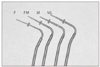
Figure 2
The images were scanned when plugger was inserted into shaped canals. Measurement of the insertion depth of the plugger (arrow line) was done by image analysis program (Image-Pro Plus, Media Cybernetic, USA).
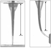
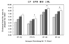
Figure 3
Insertion depth of Buchanan plugger in the straight canals. There is statistically significant difference among the pluggers in the same experimental group except the indication (*) (p < 0.05).
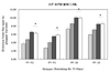
Figure 4
Insertion depth of Buchanan plugger in the straight canals. There is statistically significant difference among the experimental groups in the same plugger except the indication (*) (p < 0.05).
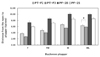
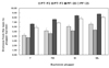
Figure 5
Insertion depth of Buchanan plugger in the curved canals. There is statistically significant difference among the pluggers in the same experimental group except the indication (*) (p < 0.05).


References
1. Seltzer S, Bender IB. The interrelationship of pulp and periodontal disease. Oral Surg Oral Med Oral Pathol. 1963. 16:1474–1490.

2. Langeland K, Rodriques H, Dowden W. Periodontal disease, bacteria, and pulpal histopathology. Oral Surg Oral Med Oral Pathol. 1974. 37:257–270.
3. Nicholls E. Lateral radicular disease due to lateral branching of the root canal. Oral Surg Oral Med Oral Pathol. 1963. 16:839–845.
4. Weine FS. The enigma of the lateral canal. Dent Clin North Am. 1984. 28:833–852.
5. Schilder H. Filling root canals in three dimensions. Dent Clin North Am. 1967. 11. 723–744.
6. Allison DA, Weber CR, Walton RE. The influence of the method of canal preparaion on the quality of apical and coronal obturation. J Endod. 1979. 5:298–304.
7. Lugassy AA, Yee F. Root canal obturation with gutta-percha: a scanning electron microscope comparison of vertical compaction and automated thermatic condensation. J Endod. 1982. 8:120–125.
8. Buchanan LS. The Continuous wave of obturation technique: 'centered' condensation of warm gutta percha in 12 seconds. Dent Today. 1996. 15:60–67.
9. Buchanan LS. The Continuous wave of condensation technique: a convergence of conceptual and procedural advances in obturation. Dent Today. 1994. 13:80–85.
10. Buchanan LS. Continuous wave of condensation technique. Endod Prac. 1998. 1:7–18.
11. Calberson FL, Deroose CA, Hommez GM, Raes H, De Moor RJ. Shaping ability of GTTM Rotary Files in simulated resin root canals. Int Endod J. 2002. 35:607–614.
12. Eldeeb ME, Boraas JC. The effect of different files on the preparation shape of severely curved canals. Int Endod J. 1985. 18:1–7.
13. Kum KY, Spangberg L, Cha BY, Jung IY, Lee SJ, Lee CY. Shaping ability of three ProFile rotary instrumentation techniques in simulated resin root canals. J Endod. 2000. 26:719–723.
14. Dummer PM, Alodeh MH. A method for the construction of simulated root canals in clear resin blocks. Int Endod J. 1991. 24:63–66.
15. Thompson SA, Dummer PMH. Shaping ability of ProFile.04 taper series 29 rotary nickel-titanium instruments in simulated root canals. Part 1. Int Endod J. 1997. 30:1–7.
16. Kazemi RB, Stennman E, Spangberg LS. Machining effciency and wear resistance of nickel-titanium endodontic files. Oral Surg Oral Med Oral Pathol Oral Radiol Endod. 1996. 81:596–602.
17. Budd CS, Weller RN, Kulild JC. A comparison of thermoplasticized injectable gutta-percha obturation techniques. J Endod. 1991. 17:260–264.
18. Smith RS, Weller RN, Loushine RJ, Kimbrough WF. Effect of varying the depth of heat application on the adaptability of gutta-percha during warm vertical compaction. J Endod. 2000. 26:668–672.
19. Guess GM, Edwards KR, Yang ML, Iqbal MK, Kim S. Analysis of Continuous-wave obturation using a single-cone and hybrid technique. J Endod. 2003. 29:509–512.
20. Bowman CJ, Baumgartner JC. Gutta-percha obturation of lateral grooves and depressions. J Endod. 2002. 28:220–223.
21. Peters OA, Peter CI, Schonenberger K, Barbaknow F. ProTaper rotary root canal preparation: effects of canal anatomy on final shape analysed by micro CT. Int Endod J. 2003. 36:86–92.
22. Lee CH, Cho KM, Hong CU. Effect of various canal preparation techniques using rotary nickel-titanium files on the maintenance of canal curvature. J Korean Acad Conserv Dent. 2003. 28:41–49.
23. Yun HH, Kim SK. A comparison of the shaping abilities of 4 nickel-titanium rotary instruments in simulated root canals. Oral Surg Oral Med Oral Pathol Oral Radiol Endod. 2003. 95:228–233.
24. Schneider SW. A comparison of canal prepararion in straight and curved canals. Oral Surg Oral Med Oral Pathol. 1971. 32:271–275.
 XML Download