

 PDF
PDF ePub
ePub Citation
 Print
Print


I. INTRODUCTION
The smear layer was first described by McComb in a scanning electron microscope (SEM) study1). The smear layer consists of organic and inorganic substances, including fragments of odontoblastic processes, dentinal shaving, microorganisms, and necrotic materials. Presence of this smear layer prevents penetration of intracanal medication into the irregularities of the root canal system and the dentinal tubules and also prevents complete adaptation of obturation materials to the prepared root canal surfaces2). Its removal calls for a combination of irrigating solutions as, presently, one solution cannot dissolve both organic and inorganic debris3). Upon the completion of instrumentation, a high volume final flush with ethylene diamine tetra-acetic acid (EDTA) solution followed by NaOCl was found to be most effective in removing superficial debris and smear layer from root canal walls4).
Studies5,6) have shown a significant increase in adhesive strength and resistance to microleakage of AH26 sealer when the smear layer was removed. Gettleman et al.5) reported that they did not find any changes in adhesive strengths when Sultan and Sealapex sealers were evaluated with or without the smear layer intact. Several investigators6-12) have shown less dye leakage after removal of the smear layer, with various obturation techniques and root canal sealers.
Other investigators have reported that the removal of the smear layer did not have any significant effect on microleakage of root canals using various sealers and obturation techniques13-16).
In contrast to these findings Timpawat et al.17-18) have reported that removal of the smear layer have adverse effects on microleakage of filled root canals.
A mixture of a tetracycline isomer, an acid, and detergent (MTAD) is known as an effective solution for the removal of the smear layer and does not significantly change the structure of the dentinal tubules when canals are irrigated with sodium hypochlorite and followed with a final rinse of MTAD19).
The purpose of this study was to evaluate the effect of newly developed endodontic root canal cleanser on the apical leakage of obturated root canal using an electrochemical method.
II. MATERIALS AND METHODS
Sixty non-carious human mature extracted maxillary or mandibular anterior teeth or premolars with a single root canal and a closed apex were distributed randomly into five groups of 12 teeth each. After preparing a conventional access preparation for each tooth, a K-type file (size 10 or 15) was used to determine the working length by penetrating the apical foramen and pulling back into the clinical apical foramen. Whilst cleaning and shaping up to a size 40 master apical file with a crown-down technique and Rotary 0.04 and 0.06 Taper Profile (Profile, Dentsply-Maillefer, Switzerland), the root canals were irrigated with 2 ml of 5.25% or 1.3% NaOCl in each group between each file size. 17% EDTA(REDTA, Roth International Ldt, Chicago, USA), 5.25% NaOCl or MTAD were used as a canal wall treatment. A total of 10 ml of irrigant was used in each root canal. The irrigant was delivered with a 27 gauge plastic needle (Capillary Tips, Ultradent product Inc., South Jordan, USA) reaching to within 1-2 mm from the working length in each canal.
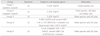
5.25% NaOCl was used as a canal irrigant in twelve teeth. After canal shaping and cleaning, root canals were obturated with only gutta-percha. These teeth were used as Group 1 (G1) or positive control group. In Group 2 (G2) or negative control group, root canals were also irrigated with 5.25% NaOCl and obturated with gutta-percha and AH plus. After that, apical portion of the roots was sealed with sticky wax. In Group 3 (G3), only 5.25% NaOCl was used as a canal irrigant, canal wall treatment and final rinse. In Group 4 (G4), specimens were irrigated with 5.25% NaOCl, treated with 5 ml of 17% EDTA for 5 minutes and final rinsed with 5.25% NaOCl. Specimens of Group 5 (G5) were irrigated with 1.3% NaOCl and treated with 5 ml of MTAD for 5 minutes. Summary of the use of canal irrigation solution was presented in table 1.
All root canals were dried with paper points and obtuated with gutta-percha and AH plus (AH plus, Dentsply, York, USA) as a sealer using a continuous wave of condensation technique except in the group 1. All roots were stored at 100% humidity for 1 day.
For all the specimens, the coronal gutta-percha was removed with #2 or #3 Gate Glidden burs and the apical 8 mm of root canal filling was left. This was confirmed radiographically. Copper wire was inserted into the canals as experimental electrodes in contact with the remaining gutta-percha. They were secured in place with sticky wax. The roots were coated with a triple layer of nailpolish except apical 8 mm in group 1, 3, 4 and 5. In the group 2, after root canal obturation, apical portion of root was covered with sticky wax and nail varnish was coated over all tooth surfaces including the sticky wax three times.
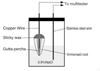
All the specimens were placed in the bottles. The copper wire as an experimental electrode was fixed in the canal and extended to the outside of the bottle cap. The stainless steel wire as a standard electrode was located at the center of the bottle as like in Park et al's study20) (Figure 1). A 0.9% NaCl solution as an electrolyte was placed in the bottle until the apical 8 mm of the roots was covered.
The electrical resistance between the standard and experimental electrode was measured over a period of 10 days using a multitester (Radio-Shack, Digital Multimeter, USA). On the first days, the electric resistance was measured at baseline and then at 0.5, 1, 2, 4, 6, 9 and 12 hour. The resistance was then measured at 24 hour and then once a day for 10 days.
A Kruskal-Wallis one-way analysis method was used along with the Student-Newman-Keuls method for mutilple comparison procedures.
III. RESULTS
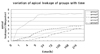
Increasing apical leakage with time was observed for all the groups.
Group 4 and 5 showed lower apical leakage than group 3 but there were no statistically significant differences between the group 3, 4 and 5 at any measurement time.
IV. DISCUSSION
Kennedy et al.7) reported that removal of smear layer reduced apical leakage when using chloroform-softened master cones with sealer and a lateral condensation technique. They described that several factors such as improved mechanical locking of sealer into patent tubules, better adhesion to cleaner canal walls, and greater canal wall sealing surface area might be responsible for this reduced apical leakage. In this study, the apical leakage of group 5 was lowest except group 2 (negative control group). Group 4 and 5 had less apical leakage than group 3. But differences between the group 3, 4 and 5 were not statistically significant. Based on the above results, the removal of smear layer was not thought to affect the apical leakage and MTAD may have better effect on the apical leakage than EDTA. In Karagoz-Kucukay's study11), comparison of the groups with smear layer present versus those with smear layer removed showed a highly significant difference, with the incidence of leakage reduced in the absence of the smear layer. In this study, there was no statistically significant difference between the group with smear layer and the groups without smear layer in the aspect of quantitative measurement.
Baumgartner et al.21) evaluated the amount of superficial debris and the smear layer that remained on the canal wall following a combination of NaOCl and 50% citric acid. They showed that citric acid or a combination of NaOCl and citric acid irrespective of the order in which they were used was more effective than NaOCl alone to remove the smear layer from the surface of instrumented canals. Because high concentrations of NaOCl are more toxic than diluted solutions and there are no significant differences between the ability of 1.3%, 2.6% and 5.25% as root canal irrigants and MTAD as a final rinse to remove the smear layer22), 1.3% NaOCl during instrumentation and MTAD as a final rinse was used to remove the smear layer in Group 5.
In this study, leakage analysis was done for 10 days. Delivanis et al.23) have shown in an electrochemical study, that there was a very sharp increase in the penetration during the first 10 days. A possible reason for this increase may be that the materials set and become dimensionally stable because of their hygroscopic and physical characteristics. Various factors such as anatomical variations, preparation of teeth, sealers used, immersion times and methods of evaluating leakage may affect the results and may be the reason for the lack of correlation between similar studies.
Further studies are necessary to evaluate the long-term apical leakage change and the coronal leakage change.
 XML Download