

 PDF
PDF ePub
ePub Citation
 Print
Print


I. INTRODUCTION
The aim of root canal preparation is to clean and shape the root canal system while maintaining the original configuration. However, traditional hand instruments often failed in achieving these objectives, especially when used in severely curved canals1,2). Over the years, many nickel-titanium rotary instruments have been developed to improve root canal preparation. More flexible files made of nickel-titanium have been effective in minimizing complications in severely curved canal preparation3,4). They are available in various designs that differ in tip and taper design, rake angles, helical angles, pitch, and presence of radial lands. Recently, new Ni-Ti rotary instrument with stainless-steel orifice shaper and quasi-rectangular cross sectional shape, NRT (Mani, Nakaakutsu, Japan) was introduced.
The purpose of this study is to determine the shaping ability of newly marketed NRT instruments in comparison with the 3 existing instruments, ProFile (Dentsply-Maillefer, Ballaigues, Switzerland), K3 (SybronEndo, West Collins, CA, USA), ProTaper (Dentsply-Maillefer, Ballaigues, Switzerland) in simulated root canals.
II. MATERIALS AND METHODS
A total of forty simulated curved root canals in clear resin blocks (Dentsply-Maillefer, Ballaigues, Switzerland) were used for this study. An apical foramen size of 0.15 mm was confirmed, and each canal had a mean canal length of 17 mm. The blocks were divided into 4 groups according to the instruments used: ProFile, K3, ProTaper and NRT. Aqueous red ink was injected into the canals to enhance the image contrast. These resin blocks were scanned in a reproducible position with a scanner, and then the image data were stored in a computer. Before the Ni-Ti files were used, the canals were explored with stainless steel No.10 and 15 hand K-files until the tip was visible at the apical foramen. The working lengths were established to apical foramen.
Preparation of simulated canals
Group 1 (ProFile) was instrumented in a crown-down manner at a constant speed of 300 rpm with ProFile .06 taper instruments. Orifice Shapers 3 and 2 were used for coronal shaping until resistance was encountered. A variable tip sequence was used in a crown-down sequence until the canal was prepared to an instrument size 30 at 0.06 taper canal terminus. In group 2 (K3), the canals were prepared with K3 .06 taper files in the same manner as group 1 to a file size 30 at a .06 taper instruments. In group 3 (ProTaper), a S1 shaping file was used first and moved apically to 2 mm short of the working length. SX files were then used until resistance was encountered, followed by S1 and S2 to the working length for the shaping of the coronal two thirds of the canal. The apical portion was finished by using F1, F2 sequentially to the working length. F3 file was not used. In group 4 (NRT), the canals were prepared with NRT .06 taper instrument. SS files were used to enlarge the canal orifices and .06 NiTi files used to prepare the mid-root and apical portions of the canals to an instrument size 30 at .06 taper canal terminus.
Assessment of canal preparation
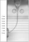
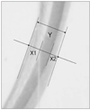
All photographs were scanned into the computer and processed by an examiner who was blinded with respect of all to the experimental groups. Using Photoshop 6.0 program (Adobe System Inc., USA), the post-operative canal images were superimposed over the pre-operative images by superposing the three dots selected in the pre- and post-instrumented images at the same positions. The change of canal curvature was measured by the Schneider method5). The ability of instruments to remain centered in the canal was determined by calculating a centering ratio after perpendicular lines were made from the canal axes at 1-, 2-, 3-, 4-, 5-, 6-, and 7-mm levels from the working length with Image analyzer (Figure 1). Observing and measuring the original canals position in the canals instrumented, the centering ratio was calculated by the formula (X1-X2)/Y [X1 represents the maximum extent of canal movement in one direction and X2 is the movement in the opposite direction. Y is the diameter of the final canal preparation]6) (Figure 2).
Recording, storage, and analysis of data
All data were stored on PC from the image processing software directly to a database file. Following error and range checks, the data were analyzed using SPSS, statistical analysis program. Change of canal curvature and differences of centering ratio at the 7 measuring points were statistically analyzed using One-way ANOVA followed by Duncan's test.
III. RESULTS
Change in canal curvature
A greater decrease in curvature was observed in the ProTaper group than in the other groups. However, there was no statistical difference (p > 0.05, Table 1).
Change in centering ratio of canals
The ability of instruments to remain centered in prepared canals at 1- and 2-mm levels was significantly better in ProFile groups than in other groups (p < 0.05). K3, NRT, and ProTaper groups presented similar change in centering ratio at 1-, 2-, 3-, 4-mm levels (p > 0.05). The change of centering ratio in NRT groups at 5-mm level was significantly greater than ProFile group and at 6- and 7-mm level than all other groups (p < 0.05, Table 2).
IV. DISCUSSION
The aim of this study was to determine the shaping ability of NRT instruments in comparison with the 3 existing instruments in simulated root canals under controlled laboratory conditions.
The centering ratio can define the ability of instruments to remain centered in shaped canals. According to the formula, the centering ratio approaches zero as X1 and X2 become closer. The lower the score, the better the instruments centered in the canal. In this present study, the results of the centering ratio in four groups at 1-, 2-mm levels indicated that the ability of instruments to remain centered in prepared canals was significantly better in Profile groups than in other groups. The ProFile instrument family, including ProFile .04 and .06 taper, Orifice Shapers, all have the same cross-sectional geometry, which is made by machining three equally spaced U-shaped grooves around the shaft of a taper Ni-Ti wire. There is a central parallel core inside that may account for the enhanced flexibility compared with Quantec7) and ProTaper8).
It was shown that 3 groups except ProFile group presented similar change in centering ratio at 1-, 2-, 3-, 4-mm levels. In previous studies it was suggested that ProTaper instruments caused greater widening of canals and lessened the canal curvature compared to other instruments9,10). In most of these studies, however, all finishing files including F3 were used to working length. All three finishing files have a fixed taper in the first 3 mm from D0 to D3. F1 has a taper of 7%, F2 has a taper of 8%, and F3 has a taper of 9% in this region. Over the remaining length of the cutting blade, a reverse taper can be found. The diameter of F3 file at 3-mm level is then 0.57 mm and it is much greater than that of .06 tapered #30 instruments. Yoshimine et al9) suggested that .06 tapered files could be used for apical preparation without creating severe aberrations, if smaller, less-tapered files were used before the use of .06 tapered ones. On the other hand, the ProTaper group showed a tendency toward apical irregularities, such as ledge and zip, despite the single slight-pressure-and-withdrawal motion of a finishing file at the end-point of preparation after instrumentation using F3 file. In this respect, F3 file was not used to avoid possible canal deviation in the present study. Guelzow et al11) reported that six rotary nickel-titanium systems including K3 and ProTaper instruments maintained the original canal curvature well with minor mean degrees of straightening. Iqbal et al12) concluded that both ProTaper and ProFile instruments are comparable to each other in regards to their ability to optimally enlarge root canal with minimal transportation and loss of working length in vitro.
It was also shown that the change of centering ratio in NRT groups at 5-, 6-, and 7-mm level was significantly greater than other groups. In the simulated resin blocks used in this study 5-mm level fell under beginning of curvature and the files for coronal shaping such as Orifice shaper in ProFile, Orifice opener in K3, and SS file in NRT were used up to this point.
The NRT files consist of the stainless-steel files used to enlarge the root canal orifices and Ni-Ti files used to prepare the mid-root and apical portions of the canal space for the continuous taper preparation. Weine13) found that stainless steel files tended to straighten curved canals. Davis et al14) reported that working length decreased as a result of canal preparation and the mean decrease in working length was significantly greater for the stainless steel group (SS hand files + Gates Glidden drills) than for the Ni-Ti group. They speculated that the differences found between stainless steel and Ni-Ti instruments are due to the superior ability of Ni-Ti to remain centered within the canal during instrumentation, thus minimizing the straightening of the canal.
The cross-section of the NRT Ni-Ti files is a Quasi-rectangle. It is suggested that this shape allows for improved file flexibility and improved cutting ability. It is also asserted that there are less possibilities for pushing debris toward root apex and danger of forcing them out of the apical foramen can be minimized with their deep space between cutting blades. However, this instrument system adopted stainless-steel as a file for preparing coronal part of root canal. In this respect, we can explain the reason why the change of canal configuration in this part was significantly greater in NRT group than in other groups.
To assess instrumentation of curved canals, clear resin blocks were used in this study. These were chosen because shape, size, taper, and curvature of the experimental canals are standardized. The credibility of resin blocks as an ideal experimental model for the analysis of endodontic preparation and preparation techniques has been validated by Weine et al.13) and Dummer et al.15). However there are limitations with the model, such as the different hardness between resin and dentin, and care should be exercised in the extrapolation of the present results to the use of these instruments in the clinical situation. Nevertheless, the use of simulated canals in resin blocks results in the opportunity to standardized the research method and to exclude parameters that could influence the preparation outcome.
V. CONCLUSION
The results indicate that ProFile system is more suitable than other instruments in the apical preparation of canals with a complicated curvature. Although the NRT system was comparable to other systems in regards to its ability to maintain the canal configuration of apical portion, this system was more influenced by the mid-root curvature due to its stainless-steel files for coronal preflaring.


 XML Download