

 PDF
PDF ePub
ePub Citation
 Print
Print


Abstract
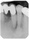
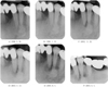
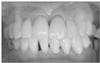
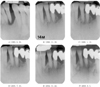
Periapical cemental dysplasia(PCD) is a condition most commonly seen in the mandibular incisor region. Radiographically it passes through the three phases(osteolytic stage, intermediate stage, and mature stage). At osteolytic stage, the lesion is similar to features associated with granuloma or cyst that arise following pulpal necrosis. So, it is important to confirm the vitality of the pulp to diagnosis.
In this case, it is difficult to confirm the vitality of involved tooth because the tooth was covered with PFG bridge. And it is unusual that the PCD lesion at mandibular incisors has occurred at first and the lesion of mandibular canine and mandibular premolar were occurred afterward.
Figures and Tables
References
1. Forget AM. Dental anomalies and their influence upon the production of diseases of the maxillary bones. Dent Cosmos. 1860. 1:342.
2. Gottlieb B. Zementexostosen, schmelztropfen und epithelnester. Z Stomatol. 1921. 19:515.
3. Hamner JE III, Scofield HH, Cornyn J. Benign fibroosseous jaw lesions of periodontal membrane origin: an analysis of 249 cases. Cancer. 1968. 22:861–878.

4. Waldron CA, Giansanti JS. Benign fibro-osseous lesions of the jaws: a clinical radiographic histologic review of sixty five cases. Part II. Benign fibro-osseous lesions of periodontal ligament origin. Oral Surg Oral Med Oral Pathol. 1973. 35:340–350.
5. Neville BW. Oral and maxillofacial pathology. 1995. 1st ed. W.B. Saunders C;460–465.
6. Stafne EC. Periapical osteofibrosis with formation of cementoma. J Am Dent Assoc. 1934. 21:1822–1829.
7. Neville BW, Albenesius RJ. The prevalence og benin fibro-osseous lesions of periodontal ligament origin black women: A radiograohic survey. Oral Surg Oral Med Oral Pathol. 1986. 62:340–344.
8. Thoma KH. Oral pathology. 1954. 4th ed. St Louis: CV Mosby C;1201–1209.
9. Chaudry AP, Spink JH, Gorlin RJ. Periapical fibrous dysplasia(cementoma). J Oral Surg. 1958. 16:483–488.
10. Scannell JM. Cementoma. Oral Surg. 1949. 2:1169–1180.
11. Zegarelli EV, Kutscher A II. The cementoma. A study of 230 patients with 435 cementomas. Oral Surg Oral Med Oral Pathol. 1964. 17:219–224.
12. Wilcox LR, Walton RE. A case of mistaken identity: periapical cemental dysplasia in an endodontically treated teeth. Endod Dent Traumatol. 1989. 5:298–301.
13. Smith S, Patel K, Hoskinson AE. Periapical cemental dysplasia: a case of misdiagnisos. Br Dent J. 1998. 185:122–123.
 XML Download