

 PDF
PDF ePub
ePub Citation
 Print
Print


Abstract
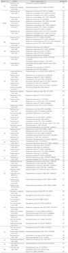
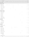
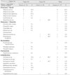
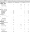
The aim of this study was to identify the bacteria isolated from acute endodontic lesions by cell culture and 16S rDNA sequencing. The necrotic pulpal tissue was collected from 17 infected root canals, which were diagnosed as being either an acute pulpitis or acute periapical abscess. Samples were collected aseptically from the infected pulpal tissue of the infected root canals using a barbed broach and a paper point. The cut barbed broaches and paper points were transferred to an eppendorf tube containing 500 ul of 1 X PBS. The sample solution was briefly mixed and plated onto a BHI-agar plate containing 5% sheep blood. The agar plates were incubated in a 37℃ anaerobic chamber for 7 days. The bacteria growing on the agar plate were identified by 16S rRNA coding gene (rDNA) cloning and sequencing at the species level. Among the 71 colonies grown on the agar plates, 56 strains survived and were identified. In dental caries involving the root canals, Streptococcus spp. were mainly isolated. Actinomyces, Clostridia, Bacteroides and Fusobacteria were isolated in the periapical lesion without dental caries. Interestingly, two new Actinomyces spp. (ChDC B639 and ChDC B631) were isolated in this study. These results showed that there was diversity among the species in endodontic lesions. This suggests that an endodontic infection is a mixed infection with a polymicrobial etiology. These results may offer the bacterial strains for pathogenesis studies related to an endodontic infection.
Figures and Tables
References
1. Gomes BP, Lilley JD, Drucker DB. Clinical significance of dental root canal microflora. J Dent. 1996. 24:47–55.

2. Fouad AF, Kum KY, Clawson ML, Barry J, Abenoja C, Zhu Q, Caimano M, Radolf JD. Molecular characterization of the presence of Eubacterium spp. and Streptococcus spp. in endodontic infections. Oral Microbiol Immunol. 2003. 18:249–255.
3. Kurihara H, Kobayashi Y, Francisco IA, Isoshima O, Nagai A, Murayama YA. Microbiological and immunological study of endodontic-periodontic lesions. J Endod. 1995. 21:617–621.
4. Abou-Rass M, Bogen G. Microorganisms in closed periapical lesions. Int Endod J. 1998. 31:39–47.
5. Baumgartner JC, Watkins BJ, Bae KS, Xia T. Association of black-pigmented bacteria with endodontic infections. J Endod. 1999. 25:413–415.
6. Rolph HJ, Lennon A, Riggio MP, Saunders WP, MacKenzie D, Coldero L, Bagg J. Molecular identification of microorganisms from endodontic infections. J Clin Microbiol. 2001. 39:3282–3289.
7. Tanner A, Stillman N. Oral and dental infections with anaerobic bacteria: clinical features, predominant pathogens, and treatment. Clin Infect Dis. 1993. 16:S304–S309.
8. Siqueira JF Jr, Rocas IN, Souto R, de Uzeda M, Colombo AP. Checkerboard DNA-DNA hybridization analysis of endodontic infections. Oral Surg Oral Med Oral Pathol Oral Radiol Endod. 2000. 89:744–748.
9. Baumgartner JC, Bae KS, Xia T, Whitt J, David LL. Sodium dodecyl sulfate-polyacrylamide gel electrophoresis and polymerase chain reaction for differentiation of Prevotella intermedia and Prevotella nigrescens. J Endod. 1999. 25:324–328.
10. Siqueira JF Jr, Rocas IN, Oliveira JC, Santos KR. Molecular detection of black-pigmented bacteria in infections of endodontic origin. J Endod. 2001. 27:563–566.
11. Maidak BL, Olsen GJ, Larsen N, Overbeek R, McCaughey MJ, Woese CR. The ribosomal database project (RDP). Nucleic Acids Res. 1996. 24:82–85.
12. Munson MA, Pitt-Ford T, Chong B, Weightman A, Wade WG. Molecular and cultural analysis of the microflora associated with endodontic infections. J Dent Res. 2002. 81:761–766. Erratum in: J Dent Res. 2003. 82:69. J Dent Res 2003. 82:247, 2002.
13. Yoo SY, Kim MK, Kim HS, Hwang HK, Kim PS, Lim SY, Oh SH, Min JB, Kook JK. Identification of bacteria from periapical abscess using 16S rDNA clone libraries. Korean J Microbiol Biotechnol. 2004. 32:195–198.
14. Sundqvist G. Taxonomy, ecology, and pathogenicity of the root canal flora. Oral Surg Oral Med Oral Pathol. 1994. 78:522–530.
15. Le Goff A, Bunetel L, Mouton C, Bonnaure-Mallet M. Evaluation of root canal bacteria and their antimicrobial susceptibility in teeth with necrotic pulp. Oral Microbiol Immunol. 1997. 12:318–322.
16. Love RM, Jenkinson HF. Invasion of dentinal tubules by oral bacteria. Crit Rev Oral Biol Med. 2002. 13:171–183.
17. Paster BJ, Boches SK, Galvin JL, Ericson RE, Lau CN, Levanos VA, Sahasrabudhe A, Dewhirst FE. Bacterial diversity in human subgingival plaque. J Bacteriol. 2001. 183:3770–3783.
 XML Download