

 PDF
PDF ePub
ePub Citation
 Print
Print


I. Introduction
Three dimensional root canal shaping and its hermetic obturation are the major elements determine the predictability of successful endodontics. In order to facilitate the irrigation process during the root canal preparation, adequate root canal shaping is also considered as a key requirement.
Conventional endodontic files are manufactured from stainless-steel, and these files have been used successfully with the concept of step-back preparation technique. Since the end of 1980's, files made of nickel-titanium (Ni-Ti) have become available. Although hand files made of Ni-Ti are available, engine-driven rotary Ni-Ti instruments and crown-down techniques have revolutionized root canal preparations and are gaining in popularity1). Numerous studies have shown that Ni-Ti rotary instruments can effectively produce a well-tapered root canal form sufficient for obturation, with minimal risk of transporting the original canal2-5).
Many Ni-Ti file systems have been introduced to the market. Most of these Ni-Ti file systems - e.g. ProFile® (Dentsply Maillefer, Ballaigues, Switzerland), K3TM (SybronEndo, Glendora, France), Hero642® (Micromega, Besancon, France) - have a constant tapered shaft design, while these have various rake angles and radial land respectively6-8). The recently developed ProTaper® (Dentsply Maillefer, Ballaigues, Switzerland) has a progressive tapered shaft design9,10). They are manufactured in different sizes from ISO standardization and each instrument has a variable taper. ProTaper® has two types on the market, the manual ProTaper® and rotary ProTaper®. The ProFile® system can be categorized as a passive instrument, while the ProTaper® (manual and rotary) system works with an active cutting motion10).
There have been many studies suggesting that rotary preparation with Ni-Ti instruments has many advantages over hand preparation with conventional stainless-steel instruments, both for experienced and for inexperienced operators5,11,12). However, few studies13-15) have addressed the use of Ni-Ti file systems by undergraduates. Currently no curriculum from any school adequately prepares its students to understand and practically apply the Ni-Ti file systems16,17). If the Ni-Ti preparation technique is included in an undergraduate curriculum, the arising question is which system would be the most effective.
The purpose of this study was to compare and evaluate the shaping abilities of three Ni-Ti file systems (ProFile®, manual ProTaper®, rotary ProTaper®) used by undergraduate students.
II. Materials and Methods
Fifty senior undergraduate students at the Dental College of Pusan National University (Busan, Korea) with no practical experience using Ni-Ti file systems in root canal preparation participated in this study. The regular curriculum instruction in root canal shaping procedures consists of six hours of lecture and eight hours of model (extracted tooth) exercises with stainless-steel files. This was the entirety of the students exposure to the procedures. In addition to the lectures on basic and practical endodontics of the regular curriculum, the students received two hours of lecture and watched video about the three systems used in this study. Respective instrumentation procedures were demonstrated before the start of the experiment.
The students prepared 150 simulated curved root canals in resin blocks (Endo Training Bloc; Dentsply Maillefer, Ballaigues, Switzerland) using the three Ni-Ti file systems - ProFile® (PF), manual ProTaper® (MPT), rotary ProTaper® (RPT). Every student prepared three simulated root canals with each system.
1. Specimen and instrumentation
1-1. Simulated root canals
The simulated root canal blocks used in this study had a vertical length of 14 mm straightly from the level of apical foramen to orifice level and the root canals had about 16 mm of working length and about 40° curvature angle. Aqueous red ink was injected into the canals to enhance the image contrast. These resin blocks were scanned in a reproducible position with a scanner (Scanjet®; C8510A, Hewlett-Packard, California, USA), and then the image data was stored in a personal computer.
1-2. Instrumentation
All Ni-Ti files used in this study were new ones. These files were used in the manner of manufacturer's recommendation. The MPT was operated by hand. Both the PF and RPT were operated by an electric motor (Tecnika®; ATR, Pistola, Italy) set at a speed of 300 rpm and torque of 30 (Tecnika motor setting value) in a 16:1 reduction handpiece. These settings were within the range suggested by the manufacturer. The instrumentation sequence of each system is summarized in Table 1. During the procedures, all simulated canals were verified the patency, then irrigated and lubricated with the RC-prep® (Stone Pharmaceuticals, Philadelphia, USA) step-by-step.
2. Measurement techniques
2-1. Instrument distortion or breakage
After root canal preparation, the Ni-Ti files were evaluated for distortion or breakage using a microscope at 25× magnification (OPMI® pico Surgical Microscope; Carl Zeiss, Oberkochen, Germany).
2-2. Aberrations
After root canal preparation was completed, aqueous methylene blue solution was injected into the enlarged canals. The resin blocks were scanned again in a reproducible position as previously described. The scanned images were assessed on a 17 inch TFT-LCD monitor (Sync-Master® CX701N; Samsung, Suwon, Korea) using Adobe® Photoshop ver.7.0 (Adobe, San Jose, California, USA). Assessments were made according to the presence of various types of canal aberrations such as apical zip, elbow and ledge.
2-3. Preparation time
Canal preparation time was recorded in minutes and seconds by each student. The recorded time included the time for irrigation, changing instruments and recapitulation.
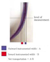
2-4. Instrumented canal width, net transportation, and centering ratio
Using Adobe® Photoshop software, the pre- and post-instrumented canal images were superimposed on one another and were observed at a magnification of 156 times. Measurements were taken horizontally at three different levels (1, 3, and 5 mm) from the level of apical foramen. The levels are selected for evaluation of terminal point, mid-point, and beginning point of root canal curvature.
Widths were measured linearly from the pre-instrumented point to the post-instrumented wall inwardly and outwardly at each level. These values were used to calculate the instrumented canal width and post-instrumented canal diameter.
The net transportation (deviation) was determined from the discrepancy between outward and inward-instrumented width (Figure 1). The direction of canal transportation was acquired by the negative or positive value. A negative value indicates an inward deviation.
The centering ratio was computed to evaluate the movement of root canal's center after preparation. The ratio was calculated using the following method: the absolute value of net transportation divided by whole width of the post-instrumented canal. The centering ratio is a measure of the ability of the instrument to stay centered in the canal; the smaller the ratio, the better the instrument remained centered in the canal9,18,19).
3. Statistical analysis
Statistical analysis of the collected data was performed with SPSS™ version 10.0 (SPSS Inc., Chicago, IL, USA). The measured data was first checked for variance homogeneity by Levene's test. When the data had normal distribution, statistical analysis was performed with ANOVA and Scheffe test for post-hoc comparison. Differences revealed in the data were designated as significant at P < 0.05. When the data did not have a normal distribution, the nonparametric Kruskal-Wallis test and Bonferroni procedure were done.
III. Results
1. Instrument distortion or breakage
There were only two broken files. One ProFile® .06 #25 and one rotary ProTaper® F1 were fractured by each student. The manual ProTaper® had no fractures and none of the three systems had other distortions or deformations.
2. Aberrations
The resin blocks instrumented with MPT or RPT had more zips and elbows than those with PF (Table 2).
3. Preparation time
The resin blocks that had procedural errors or other factors interfering with completion (such as instrument separation, ledge-not negotiated) were excluded from the samples for statistical analysis. The time needed for completion for each system is presented in Table 3. Students required more time for the entire preparation with the MPT system. The nonparametric Kruskal-Wallis test and Bonferroni procedure showed statistically significant differences between the groups (P < 0.05).
4. Instrumented canal width, net transportation, and centering ratio
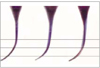
Three representative superimposed images of pre-instrumented and post-instrumented blocks are presented in Figure 2.
The instrumented canal widths in the RPT were significantly larger than those of the PF and MPT at the 1 mm and 3 mm levels. At the 5 mm level, both of the ProTaper® systems, MPT and RPT, enlarged the root canals more than PF (P < 0.05) (Table 4).
There were no significant differences between the two ProTaper® groups in net transportation. However, the root canals preparated by the two ProTaper® systems deviated more than the ProFile® system at the 1 mm and 5 mm levels (P < 0.05). Most of the deviations were inward with each of the ProTaper® systems having an outward deviations at the 1 mm level (Table 5).
The centering ratios at the 1 mm and 3 mm levels had no significant differences (P > 0.05). The centering ratio at the 5 mm level was largest in MPT and the differences between the systems were significant (P < 0.05) (Table 6).
5. Questionnaires and Preference
Students' responses are summarized in Table 7. While no students preferred MPT, however, 32 students preferred RPT over PF. Eleven students cited the length of time it took to operate the MPT as a disadvantage. Several students cited each system as being easy to handle (RPT 17, PF 7, and MPT 5). The safety about Ni-Ti file system was also mentioned. Five students indicated that they felt safe during the trial procedure using PF and one with MPT. However, RPT was not cited as feeling safe, nine students stated the anxiety about instrument breakage during use. In relation to tactile sense, nine students mentioned that MPT was more conservative than other systems. Twelve students complained that RPT often removed material aggressively and showed instrument locking. In using ProTaper® systems (MPT and RPT), six students complained that F1 and F2 sequences were more difficult to follow and resistant than other steps. Similarly 17 students mentioned that the sequence from .04 #25 to .06 #20 in PF was difficult and resistant. Four students felt that the RPT's cutting ability was better and one student felt the same about MPT.
Finally, we evaluated and compared the simulated root canals instrumented by the two preferred groups. The 31 simulated canals prepared by RPT (a student's sample was excluded because of file fracture) and 18 canals by PF were evaluated. The data was analysed by Levene's test and t-test or nonparametric Mann-Whitney test. The statistical results were not different from that of previously described.
IV. Discussion
Rotary Ni-Ti instruments are used efficiently in root canal treatments throughout the world, but not by all dentists. It may not be common for most operators to practice on extracted teeth or plastic blocks to evaluate the possibilities and limitations of a new technique, device, or instrument11). Like many clinical practitioners, most dental students do not have many opportunities to practice new techniques. The aim of this study was to evaluate the efficacy of the three Ni-Ti file systems used by undergraduates.
A survey about the use of rotary Ni-Ti instruments reported that these instruments were used by 22% of general dentists and 64% of endodontists20). The two main reasons for not using rotary Ni-Ti were 'no perceived advantage'and 'high fragileness'. The next two most common problems encountered were 'binding' and 'ledging'. On the other hand, very high proportions of positive experiences were also noted.
The main problem inherent in rotary preparation by inexperienced operators is the risk of instrument fracture. A study8) that compare the breakage and distortion of various Ni-Ti file systems in curved canals reported that the ProFile® system had statistically significantly more distortions than the ProTaper® system. However, there was no distortion and little breakage in this study. The reason might be that the torque control motors were used for PF and RPT. The low-torque control motors will reverse the rotation of the instrument when the instrument is subjected to stress levels equal to the preset torque value21). Most manufacturers suggest that the Ni-Ti instruments should be used about 10 times. The Ni-Ti files used in this study were new ones, so it is surmised that there were fewer fractures due to fatigue or over-use. Instrumentation technique, instrument design, angle and radius of canal curvature and the torque control motor all have an influence on the fracture rate of Ni-Ti files21-23). Strictly, all of these factors do not always seem to exert an influence on beginners, dissimilarly on the experienced.
Statistical analysis of root canal aberrations was not possible due to the low number of occurrences. However, the MPT or RPT had a few more zips and elbows than the PF. The reason may be over-uses of F series files in ProTaper® system which can remove tooth structure excessively when left in the canal too long with their active design24). It is therefore of utmost importance to follow the manufacturer's instructions and not to leave the RPT prepare the root canal for longer than 1 second or not to use the MPT too much when reaching the desired working length. These aberrations can be diminished with several preclinical trial uses and following the manufacture's instructions.
In the present study, the RPT took significantly less instrumentation time than the other systems. The same results were reported in other studies25,26) comparing the ProTaper® and other Ni-Ti rotary file systems. Statistically significant differences existed between the groups although there were large deviations in the amount of preparation time. It is natural that the manually controlled MPT would take longer to use. This was the reason that no student chose MPT as a preferred system. This statistically significant difference is, however, to be interpreted with caution, as the preparation times were recorded by students themselves and the time included not only actual instrumentation time but also the time for irrigation, changing instruments and recapitulation.
Manufacturers present the preservation of tactile sense as an advantage of the MPT system. However, beginners who had no experience using rotary file systems or even stainless steel files in clinical situations could not feel and did not need tactile sensation from the MPT. For this reason, they preferred the relatively fast and simple rotary systems.
Instrumented canal width simply implicates the cutting ability of the applied system. In this study, the canal width formed by MPT or RPT was significantly larger than that formed by PF. This seemed to be resulted from the thicker instrument, especially the F series files, which have .07 (F1) or .08 (F2) apical taper in the ProTaper® system. Also, this might have resulted from the active cutting feature with that both ProTaper® systems have.
The studies27) that compared apical transportation between ProFile® and ProTaper® showed no statistically significant differences. However, in present study with the viewpoint of net transportations, all root canals usually deviated inwardly at all levels with exception of 1 mm level (the terminal point of the curvature) of MPT and RPT. At this level both ProTaper® systems had outward deviations. This tendency may have resulted from the greater restoring force of the ProTaper® file on the outer curve and the diminished super-elastic property of Ni-Ti due to the thick end of the ProTaper®, especially in F series files.
In centering ratio, there were significant differences at the only level of 5 mm between the systems and PF had the better centering ability than the other two groups at 5 mm level. Nevertheless, the centering ratios were not significantly different between the groups in level 1 mm and 3 mm. The results of the centering ratio can be used as an index for the capability of the instrument to stay centered in the canal. The centering ratio was used as one of the most important qualitative evaluations of the present study. In narrow and curved root canals, the coronal canals must be enlarged to facilitate access to the apical canal and obturation. The centering ratios at the 5 mm level indicate that both ProTaper® systems did not remain centered at the measured level of canal. Nevertheless, from the viewpoint of coronal flaring and obturation facilitation, the ProTaper® system can easily provide straight access to apically and larger apical sizes.
That no student preferred MPT was beyond expectation. Students' responses indicated that the RPT cut more canal wall more quickly than the PF did, which is in keeping with the previously cited material. This suggests that the shorter working time was seen as preferential by the students. The other advantages of the RPT the students cited were its ease of handling and efficiency efficiency in cutting.
The constant tapered shaft design has more flexibility than the progressive tapered shaft design. And the U-shaped cross section is more flexible than the triangular cross section9,10). The students seemed to react to the relatively rougher feel of the ProTaper® when compared to the ProFile®.
Properly used Ni-Ti file systems enable the user to finish more predictable root canal instrumentation and limit procedural errors at the same time10). Generally, little information is available regarding the attitude of general dental practitioners towards new endodontic concepts, techniques and instruments, and on how far these have been incorporated into daily practice28). Students have less bias towards the instruments and techniques, but quickly learn to manage the instrumentation system. The practitioners, however, who have biased information about the rotary Ni-Ti instrument initially began to use these instruments aggressively. This sometimes ended in breakage and failure of the instruments11). So, it is suggesting that suitable preclinical training should be taken enough in advance of clinical practice on patients. It is advised to gain the requisite experience on 20 teeth including extracted teeth29).
In this study, the ProFile® system represented a constant tapered and passive cutting file with negative rake angle and the recently introduced ProTaper® system as the only progressive tapered file on the market represented an active cutting file. The ProTaper® system is the only product that has two systems with different usage. Based on the results, novice operators even the students can do the root canal preparation with any of the Ni-Ti systems used in this study. Although the root canals instrumented using the ProTaper® systems showed a bit more cutting from a quantitative viewpoint, in the centering ratio of qualitative aspect, there were little differences between the three systems.
Based on the comparative report of mathematical models of ProTaper® and ProFile®, the results has shown that the ProTaper® model might be more indicated for narrow canals and curved canals during the initial phase of shaping and that the ProFile® model might be more indicated for wider canals and curved canals in the final phase of shaping30). Further investigations such as hybrid techniques using various Ni-Ti file systems in the undergraduate students are needed prior to integrate the Ni-Ti file systems to regular undergraduate curriculum.
V. Conclusions
Under the condition of this study, both ProTaper® systems allowed significantly more removal of root canal wall than the ProFile® system. In the important other aspects such as the centering ratio, there was no significant differences between the systems.
Senior dental students were able to prepare curved root canals with any Ni-Ti file systems with little aberration and great conservation of tooth structure. Students want to learn effective methods and at the same time simple rotary procedures. The rotary ProTaper® systems were one of the most compatible to these students from the point of view of cutting ability. The ProFile® system was also compatible in safe and gentle shaping.


 XML Download