

 PDF
PDF ePub
ePub Citation
 Print
Print


Abstract
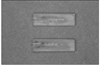
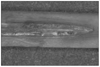
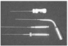
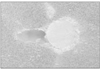
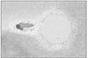
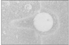
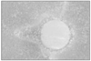
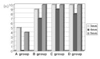
The aim of this study was to evaluate the effectiveness of sealer placement in simulated root canal extensions. Forty resin blocks were attained from the Endo-training Bloc. In each block, the simulated root canal was made with #20, 08taper GT file. After each block was longitudinally split into two halves, a standardized groove was prepared on one canal wall of two halves to simulate the canal extensions with various irregularities. The two halves of each block were assembled and all simulated root canals were obturated by single cone method with AH26 sealer. Four different methods of sealer placement were used: group A, #20 K-file; group B, ultrasonic file; group C, lentulo spiral; group D, EZ-Fill bi-directional spiral. All obturated blocks were stored in 100% humidity at 37℃ for 1 week. Using a low speed saw, each block was sectioned horizontally. Images of the sections were taken using a stereomicroscope at × 30 magnification and a digital camera. The amount of the sealer in the groove was evaluated using a scoring system, a higher score indicated better sealing effectiveness. The data was statistically analysed by Fisher's Exact Test.
The sealing score was the lowest, specially at the middle area of canal extensions in group A, and that was statistically significant difference from other groups. In conclusion, the ultrasonic file, lentulo spiral and EZ-Fill bi-directional spiral were effective methods of sealer placement in simulated canal extensions. The K file was the least effective method, specially at the middle area of canal extensions.
Figures and Tables


References
1. ElDeeb ME. The sealing ability of infection-molded thermoplasticized gutta-perch. J Endod. 1985. 11:84–86.
2. Hata G, Kawazoe S, Toda T, Weine FS. Sealing ability of Thermafil with and without sealer. J Endod. 1992. 18:322–326.

3. Fan B, Wu MK, Wesselink PR. Leakage along warm gutta-percha fillings in the apical canals of curved roots. Endod Dent Traumatol. 2000. 16:29–33.
4. Lin LM, Skriber JE, Gaengler P. Factors associated with endodontic treatment failures. J Endod. 1992. 18:625–627.
5. Hoen MM, Labounty GL, Keller DL. Ultrasonic endodontic sealer placement. J Endod. 1988. 14:169–174.
6. West WA, Labounty GL, Keller DL. Obturation quality utilizing ultrasonic cleaning and sealer placement followed by lateral condensation wth gutta percha. J Endod. 1989. 15:507–511.
7. Wiemann Ah, Wilcox LR. In vitro evaluation of four methods of sealer placement. J Endod. 1991. 17:444–447.
8. Hall MC, Clement DJ, Dove SB, Walker WA. A comparison of sealer placement techniques in curved canals. J Endod. 1996. 22:638–642.
9. Kahn FH, Rosenberg PA, Schertzer L, Korthals G, Nguyen PNT. An in-vitro evaluation of sealer placement methods. Int Endod J. 1997. 30:181–186.
10. Musikant BL, Cohen BI, Deutsch AS. Simplified obturation of tapered canal preparations. Compend Contin Educ Dent. 1998. 19:1152–1155.
11. Musikant BL, Cohen BI, Deutsch AS. Rethinking endodontics: attaining total obturation of the root canal system with a simplified system. Gen Dent. 1999. 47:73–82.
12. Seidman D. A general dentist's viewpoint of two new endodontic techiques. Compend Contin Educ Dent. 1999. 20:921–924.
13. Musikant BL, Cohen BI, Deutsch AS. Report of a simplified endodontic technique. Compend Contin Educ Dent. 1999. 20:1088–1090.
14. Barthel CR, Losche GM, Zimmer S, Roulet JF. Dye penetration in root canals filled with AH-26 in different consistencies. J Endod. 1994. 20:436–439.
15. Dalat DM, Spangberg LSW. Comparison of apical leakage in root canals obturated with various gutta-percha techniques using dye vacuum tracing method. J Endod. 1994. 20:315–319.
16. Green D. Morphology of the pulp cavity of permanent teeth. Oral Surg Oral Med Oral Pathol. 1955. 8:743–759.
17. Jeffrey IWM, Saunders WP, Thomas GE. An investigation into the movement of sealer during placement of gutta-percha points. Int Endod J. 1986. 19:21–28.
18. Wu MK, Ozok AR, Wesselink PR. Sealer distribution in root canals obturated by three techniques. Int Endod J. 2000. 33:340–345.
 XML Download