

 PDF
PDF ePub
ePub Citation
 Print
Print


Abstract
The purpose of this study was to evaluate the canal configuration after shaping by ProFile, ProTaper and K-Flexofile in simulated resin canals with different angles of curvature.
Three types of instruments were used : ProFile, ProTaper, K-Flexofile. Simulated root canals, which were made of epoxy resin, were prepared by ProFile, ProTaper with rotary instrument using a crown-down pressureless technique, and hand instrumentation was performed by K-Flexofile using a step-back technique. All simulated canals were prepared up to size 25 file at end-point of preparation. Pre and post instrumentation images were recorded with Scanner. Assessment of canal shape was completed with Image Analysis program. Measurements were made at 1, 2, 3, 4, 5, 6, 7, 8, 9 and 10 mm from the apex. At each level, outer canal width, inner canal width, total canal width, and amount of transportation from original axis were recorded. Instrument deformation and fracture were recorded. Data were analyzed by means of one-way ANOVA analysis of variance and the Sheffe's test.
The result was that ProFile and ProTaper maintain original canal shape regardless of the increase of angle of curvature than K-Flexofile. ProFile show significantly less canal transportation and maintained original canal shape better than ProTaper.
Figures and Tables
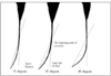 | Figure 1The three different angles of curvature (by Schneider method).
Working length was adjusted to 18 mm, the beginning point of curvature was began at 10 mm from the canal orifice.
|
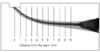 | Figure 2A diagram is indicating the points at which the canal widths were measured after superimposition of preinstrumentation and postinstrumentation images.
|
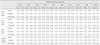
Table 1
Mean values of outer, inner, total canal width and amount of transportation in 15 degree of canal curvature (Unit: mm, Mean ± S.D.)
![]()
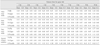
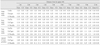
Table 2
Mean values of outer, inner, total canal width and amount of transportation in 30 degree of canal curvature (Unit: mm, Mean ± S.D.)
![]()
References
1. Schilder H. Filling root canals in three dimensions. Dent Clin North Am. 1967. 723–744.

2. Zmener O, Balbachan L. Effectiveness of nickel-titanium files for preparing curved root canals. Endod Dent Traumatol. 1995. 11:121–123.
3. Weine FS. Endodontic therapy. 1989. 4th ed. St. Louis: Mosby;277.
4. Walton R. Current concepts of canal preparation. Dent Clin North Am. 1992. 36:309–326.
5. Roane J, Sabala C, Duncanson M. The "balanced force" concept for instrumentation of curved canals. J Endod. 1985. 11:203–211.
6. Morgan LF, Montgomery S. An evaluation of the crown-down pressureless technique. J Endod. 1984. 10:491–498.
7. Chenail BL, Teplitsky PE. Endosonics in curved root canal. J Endod. 1985. 11:369–374.
8. Walsh C, Messee HH, EIDeeb ME. Effect of varying ultrasonic power setting canal preparation. J Endod. 1990. 16:273–278.
9. Ingle JI, Taintor JF. Endodontics. 1985. 3rd ed. Philadelphia: Lea & Febiger;26–37.
10. Walia H, Brantley WA, Gerstein H. An initial investigation of the bending and torsional properties of Nitinol root canal files. J Endod. 1988. 14:346–351.
11. Esposito PT, Cunningham CJ. A comparison of canal preparation with nickel-titanium and stainless steel instruments. J Endod. 1995. 21:173–176.
12. Park HS, Lee MG, Kim JJ, Lee JY. A study on the shape of a canal prepared with profiles in a curved canal. J Korean Acad Conserv Dent. 1999. 24:633–637.
13. Ko HJ, Baek SH. A study of histomorphological change of curved root canal preparation using GT rotary file, profile and stainless steel K-file. J Korean Acad Conserv Dent. 2002. 27:612–621.
14. Calberson FLG, Deroose CAJG, Hommez GMG, Raes H, De Moor RJG. Shaping ability of GT™ Rotary Files in simulated resin root canals. Int Endod J. 2002. 35:607–614.
15. ElDeeb ME, Boraas JC. The effect of different files on the preparation shape of severely curved canals. Int Endod J. 1985. 18:1–187.
16. Lim KC, Webber J. The validity of simulated root canals for the investigation of the prepared root canal shape. Int Endod J. 1985. 18:240–246.
17. Dummer PMH, Alodeh MHA, Al-Omari MAO. A method for the construction of simulated root canals in clear resin blocks. Int Endod J. 1991. 24:63–66.
18. Schneider SW. A comparison of canal prepararion in straight andcurved canals. Oral Surg. 1971. 32:271–275.
19. Yun HH, Kim SK. A comparison of the shaping abilities of 4 nickel-titanium rotary instruments in simulated root canals. Oral Surg Oral Med Oral Pathol Oral Radiol Endod. 2003. 95:228–233.
20. Hata G, Uemura M, Kato AS, Imura N, Novo NF, Toda T. A comparison of shaping ability using ProFile, GT file, and Flex-R endodontic instruments in simulated canals. J Endod. 2002. 28:316–321.
21. Bishop K, Dummer PM. A comparison of stainless steel Flexofiles and nickel-titanium NiTiFlex files during the shaping of simulated canals. Int Endod J. 1997. 30:25–34.
22. Coleman CL, Svec TA. Analysis of Ni-Ti versus stainless steel instrumentation in resin simulated canals. J Endod. 1997. 23:232–235.
23. Park H. A comparison of Greater Taper files, ProFiles, and stainless steel files to shape curved root canals. Oral Surg Oral Med Oral Pathol Oral Radiol Endod. 2001. 91:715–718.
24. Peters OA, Peter CI, Schonenberger K, Barbaknow F. ProTaper rotary root canal preparation: effects of canal anatomy on final shape analysed by micro CT. Int Endod J. 2003. 36:86–92.
25. Kavanagh D, Lumley PJ. An in vitro evaluation of canals preparation using ProFile .04 and .06 taper instruments. Endod Dent Traumatol. 1998. 14:16–20.
26. Bryant ST, Dummer PM, Pitoni C, Bourba M, Moghal S. Shaping ability of .04 and .06 taper ProFile rotary nickel-titanium instruments in simulated root canals. Int Endod J. 1999. 32:155–164.
27. Oh HJ, Hong CH, Jo YB. The effect of niti rotary instrumentation on the configuration of apical root canal. J Korean Acad Conserv Dent. 1997. 22:244–253.
28. Southard DW, Oswald RJ, Natkin E. Instrumentation of curved molar root canals with the Roan technique. J Endod. 1987. 13:479–489.
29. Kum KY, Spangberg L, Cha BY, Jung IY, Lee SJ, Lee CY. Shaping ability of three ProFile rotary instrumentation techniques in simulated resin root canals. J Endod. 2000. 26:719–723.
30. Thompson SA, Dummer PM. Shaping ability of Lightspeed rotarynickel-titanium instruments in simulated root canals. Part 1. J Endod. 1997. 23:698–702.
 XML Download