

 PDF
PDF ePub
ePub Citation
 Print
Print


ABSTRACT
The purpose of this study was to evaluate the effect of dual bonding technique by comparing micro-shear bond strength between two different luting methods of resin cement to tooth dentin. Three dentin bonding systems(All-Bond 2, One-Step, Clearfil SE Bond), two temporary cements (Propac, Freegenol) were used in this study.
In groups used conventional luting procedure, dentin surfaces were left untreated. In groups used dual bonding technique, three dentin bonding systems were applied to each dentin surface. All specimens were covered with each temporary cement. The temporary cements were removed and each group was treated using one of three different dentin bonding system. A resin cement was applied to the glass cylinder surface and the cylinder was bonded to the dentin surface. Then, micro-shear bond strength test was performed. For the evaluation of the morphology at the resin/dentin interface, SEM examination was also performed.
Conventional luting procedure showed higher micro-shear bond strengths than dual boning technique. However, there were no significant differences.
Freegenol showed higher micro-shear bond strengths than Propac, but there were no significant differences.
In groups used dual bonding technique, SE Bond showed significantly higher micro-shear bond strengths in One-Step and All-Bond 2 (p < 0.05), but there was no significant difference between One-Step and All-Bond 2.
In SEM observation, with the use of All-Bond 2 and One-Step, very long and numerous resin tags were observed. This study suggests that there were no findings that the dual bonding technique would be better than the conventional luting procedure.
REFERENCES
1. Tyas MJ, Anusavice KJ, Frencken JE, Mount GJ. Minimal intervention dentistry. Int Dent J. 20:1–12. 2000.
2. Versluis A, Douglas WH, Cross M, Sakaguchi RL. Does an incremental filling technique reduce the polymerization shrinkage stress? J Dent Res. 75:871–878. 1996.
3. Fontana M, Dunipace AJ, Gregory RL, Noblitt TW, Li Y, Park KK, Stookey GK. An in vitro microbiological model for studying secondary careis formatioon. Caries Res. 30:112–118. 1996.
4. Inokoshi S, Willems G, Van Meerbeek B, Lambrechts P, Braem M, Vanherle G. Dual-cure luting composites : Part Ⅰ: Filler particle distribution. J Oral Rehabil. 20:133–146. 1993.
5. Burrow MF, Nikaido T, Satoh M, Tagami J. Early bonding of resin cements to dentin. Oper Dent. 21:196–202. 1996.
6. Eliades G. Clinical relevance of the formulation and testing of dentine bonding systems. J Dent. 22:73–81. 1994.

7. Frankenberger R, Kramer N, Petschelt A. Technique sensitivity of dentin bonding : Effect of application mistakes on bond strength and marginal adaptation. Oper Dent. 25:324–330. 2000.
8. El-Mowafy OM, Bennergui C. Radiopacity of resin-based inlay luting cements. Oper Dent. 19:11–15. 1994.
9. Milleding P, Ortengren U, Karlsson S. Ceramic inlay systems : some clinical aspects. J Oral Rehabil. 22:571–580. 1995.
10. Watanabe I, Nakabayashi N, Pashley PH. Bonding to ground dentin by a Phenyl-P self-etching primer. J Dent Res. 73:1212–1220. 1994.
11. Xie J, Powers JM, McGuckin RS. In vitro bond strength of two adhesives to enamel and dentin under normal and contaminated conditions. Dent Mater. 9:295–299. 1993.
12. Kaneshima T, Yatani H, Kassai T, Watanabe EK, Yamashita A. The influence of blood contamination on bond strengths between dentin and adhesive resin cement. Oper Dent. 25:195–201. 2000.
13. Christensen GJ. Resin cements and post-operative sensitivity. J Am Dent Assoc. 131:1197–1199. 2000.
14. Paul SJ, Scha¨rer P. The dual bonding technique : A modified method to improve adhesive luting procedures. Int J Periodont Rest Dent. 17:537–545. 1997.
15. DeGoes MF, Nikaido T, Pereira PNR, Tagami J. Early bond strengths of dual- cured resin cement to resin-coated dentin. J Dent Res. 79:453. 2000.
16. Bertschinger C, Paul SJ, Luthy H, Scha¨rer P. Dual application of dentin bonding agents : effects on bond strengths. Am J Dent. 9:115–119. 1996.
17. Baier RE. Principles of adhesion. Oper Dent. (Supplement 5):1–9. 1992.
18. Terata R. Characterization of enamel and dentin surfaces after removal of temporary cement - study on removal of temporary cement. Dent Mater. 12:18–28. 1993.
19. Marshall SJ, Marshall GW, Harcourt JK. The influence of various cavity bases on the micro-hardness of composites. Aust Dent J. 27:291–295. 1982.
20. Millstein PL, Nathanson D. Effect of eugenol and eugenol cements on cured composite resin. J Prosthet Dent. 50:211–215. 1983.
21. Powell TL, Huget EF. Effects of cements and eugenol on properties of a visible light-cured composite. Pediatr Dent. 15:104–107. 1993.
22. Woody TL, Davis RD. The effect of eugenol containing and eugenol-free temporary cements on microleakage in resin bonded restorations. Oper Dent. 17:175–180. 1992.
23. Stangel I, Nathanson D, Hsu C. Shear strength of the composite bond to etched porcelain. J Dent Res. 66:1460–1465. 1987.
24. So¨derholm K-JM. Correlation of in vivo and in vitro performance of adhesive materials : A report of the ASC MD 156 task group on test methods for adhesion of restorative materials. Dent Mater. 7:74–83. 1991.
25. Swift EJ, Perdigao J, Heymann HO. Bonding to enamel and dentin : A brief history and state of the art. Quintessence Int. 26:95–110. 1995.
26. Sano H, Shono T, Sonoda H, Takatsu T, Ciucchi B, Carvalho R, Pashley DH. Relationship between surface area for adhesion and tensile bond strength-evaluation of a microtensile bond test. Dent Mater. 10:236–240. 1994.
27. Gwinnett AJ, Kanca J. Micromorphology of the bonded dentine interface and its relationship to bond strength. Am J Dent. 5:73–77. 1992.
28. Sano H, Takatsu T, Ciucchi B, Horner JA, Matthews WG, Pashley DH. Nanoleakage: leakage within the hybrid layer. Oper Dent. 20:18–25. 1995.
29. Cox CF. Evaluation and treatment of bacterial microleakage. Am J Dent. 7:293–295. 1994.
30. Paul SJ, Scha¨rer P. Effect of provisional cements on the shear bond strength of various dentin bonding agents. J Oral Rehabil. 24:8–14. 1997.
31. Christensen GJ. Resin cements and postoperative sensitivity. J Am Dent Assoc. 131:1197–1199. 2000.
32. Sorensen JA, Munksgaard EC, Odont D. Relative gap formation adjacent to ceramic inlays with combination of resin cements and dentine bonding agents. J Prosthet Dent. 76:472–476. 1996.
33. Magne P, Douglas WH. Porcelain veneers : dentin bonding optimization and biomimetic recovery of the crown. Int J Prosthodont. 12:111–121. 1999.
34. Kanca J. Resin bonding to wet substrates I-bonding to dentin. Quintessence Int. 23:39–41. 1992.
35. Tay FR, Gwinnett JA, Wei SH. Micromorphological spectrum from over drying to over-wetting acid conditioned dentin in water-free acetone-based single bottle primer/adhesives. Dent Mater. 12:236–244. 1996.
36. Nakabayashi N, Kojima K, Masuhara E. The promotion of adhesion by the infiltration of monomers into tooth substrates. J Biomed Mater Res. 16:265–273. 1982.
37. Ganss C, Jung M. Effect of eugenol- containing temporary cements on bond strength of composite to dentin. Oper Dent. 23:55–62. 1998.
38. Yoshiyama M, Carvalho RM, Sano H, Horner JA, Brewer PD, Pashley DH. Regional bond strengths of resins to human root dentine. J Dent. 24:435–442. 1996.
39. Pashley DH, Pashley EL. Dentin permeability and restorative dentistry : a status report for the American Journal of Dentistry. Am J Dent. 4:5–9. 1991.
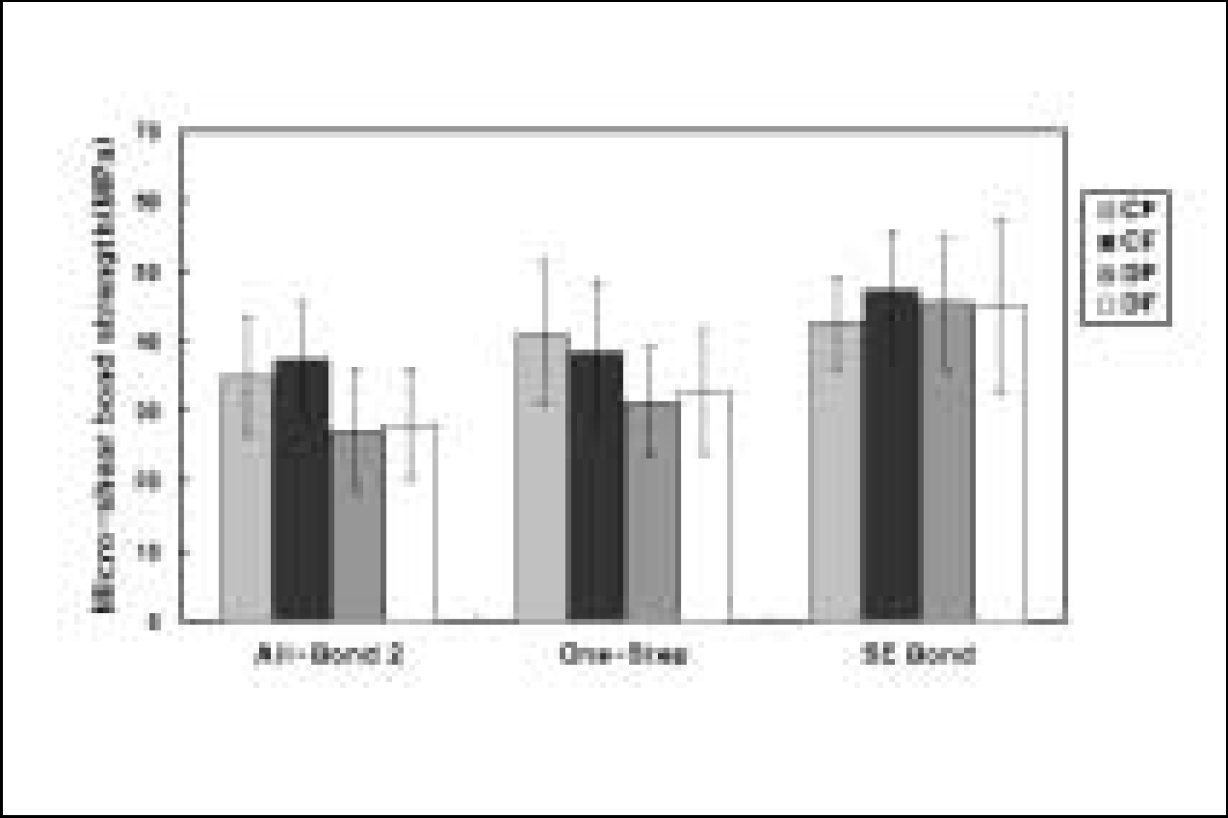
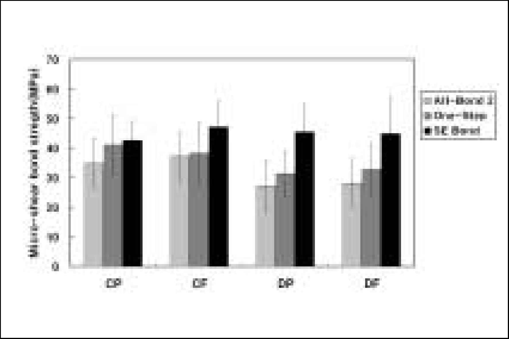
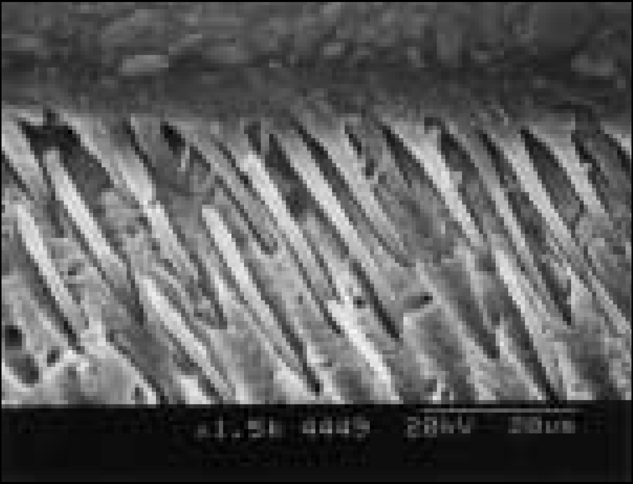
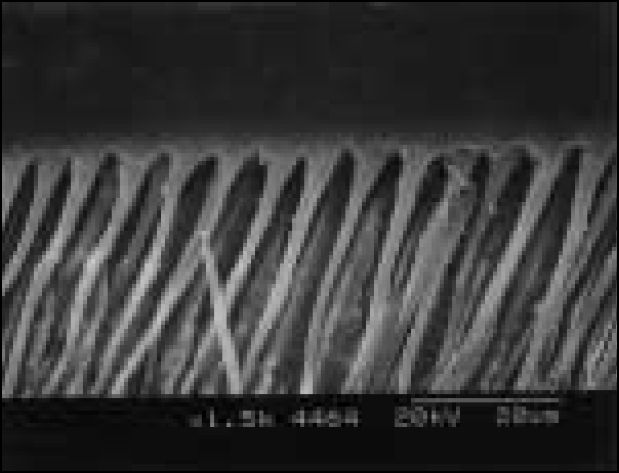
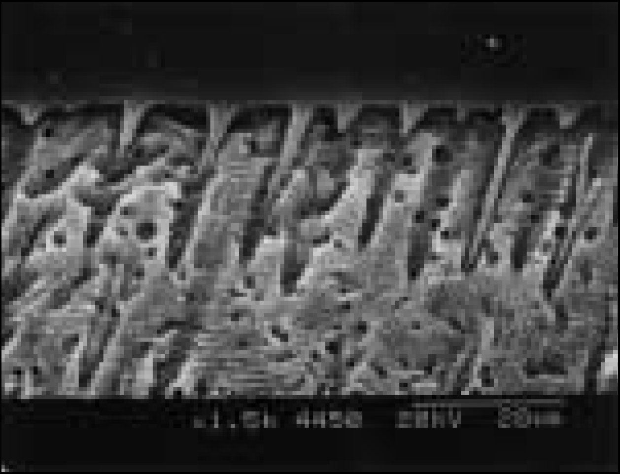
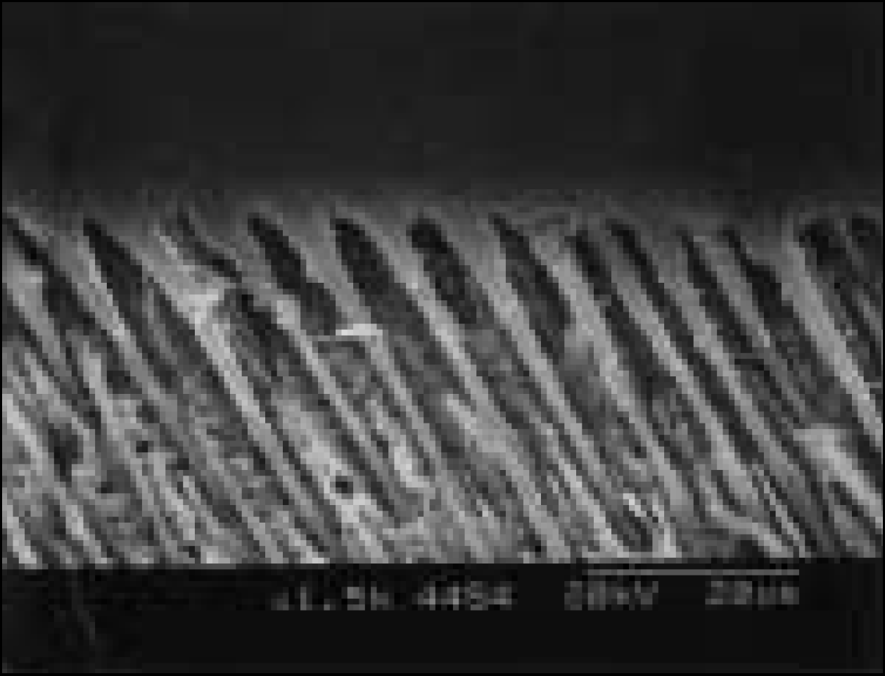

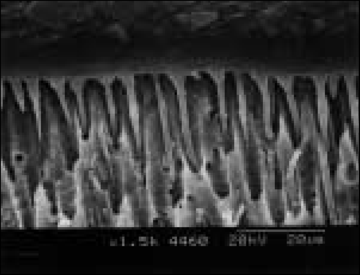
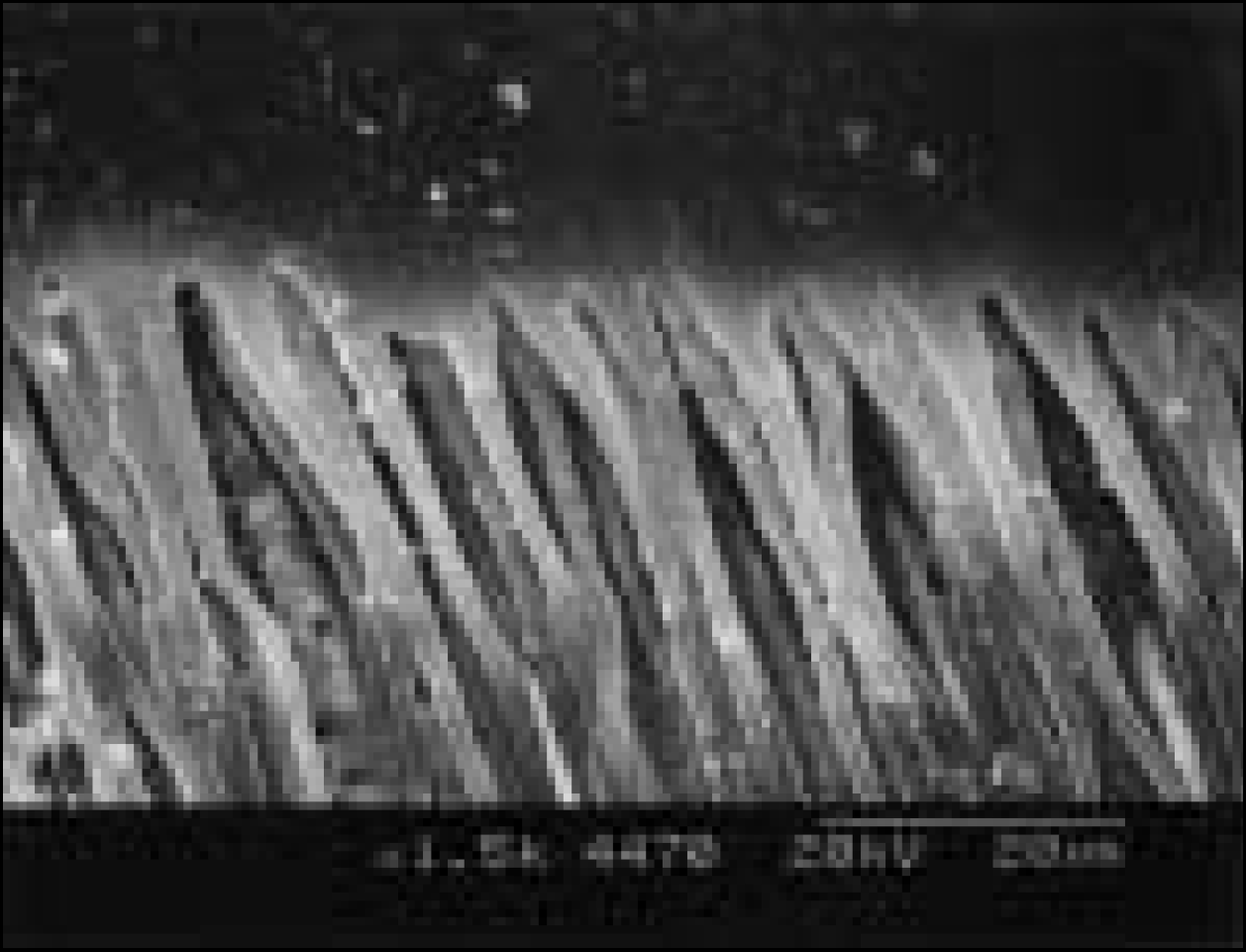
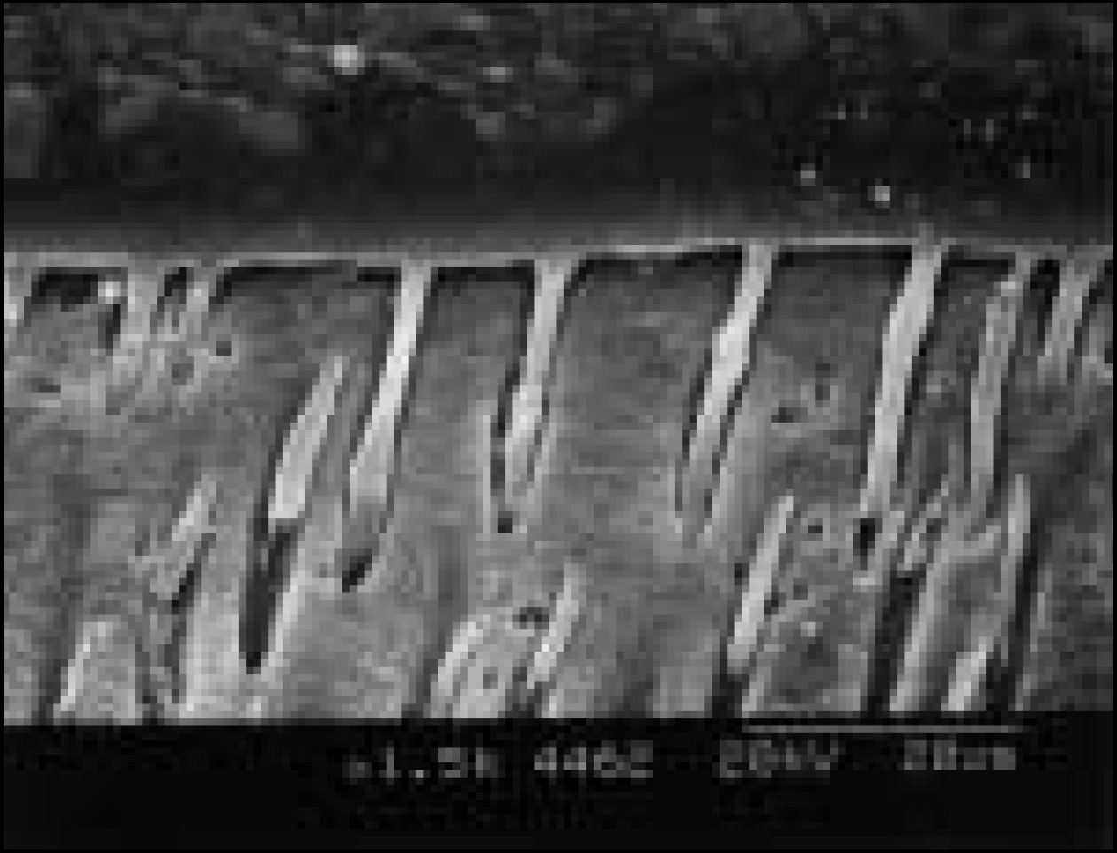
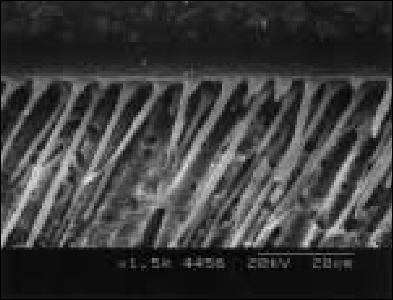
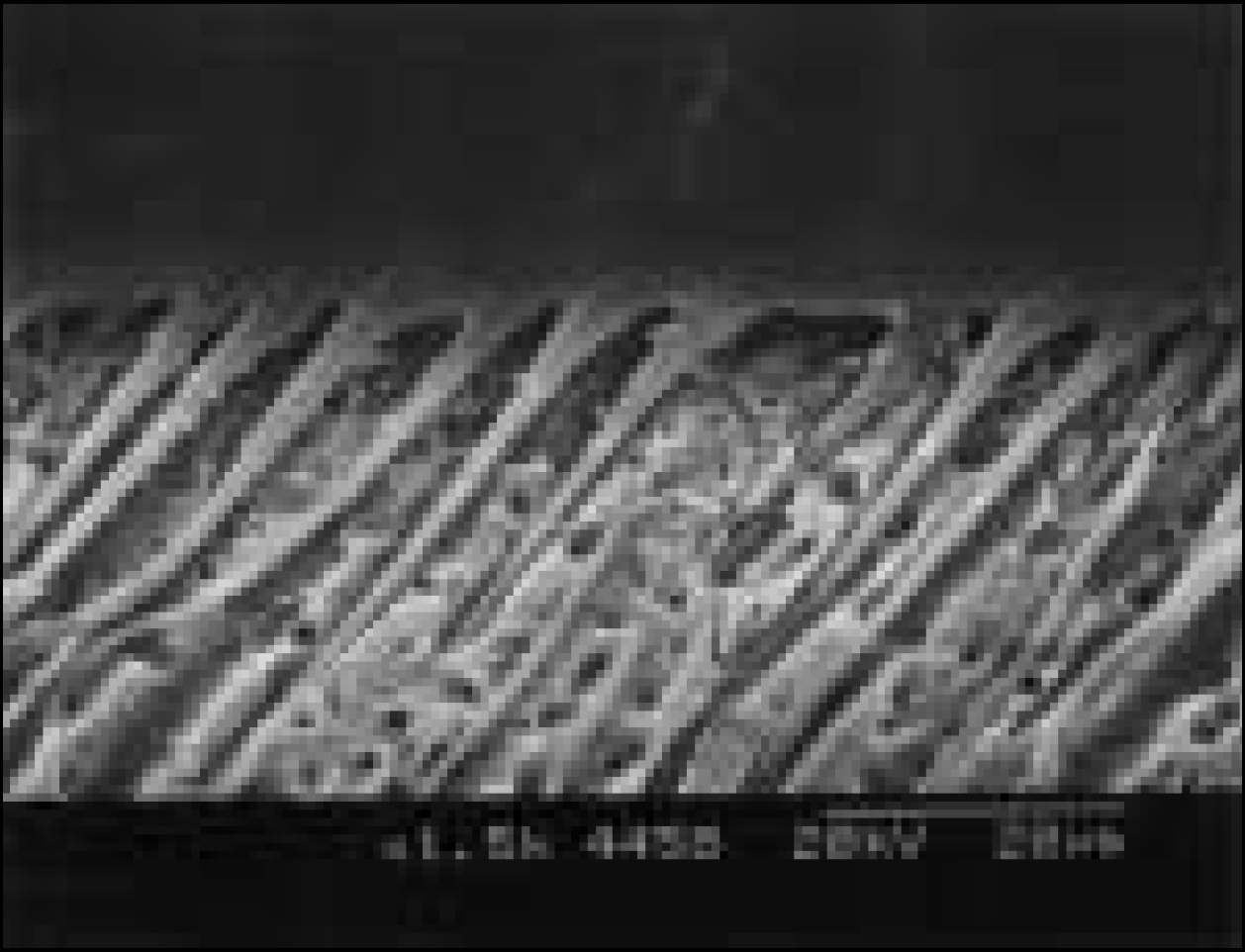
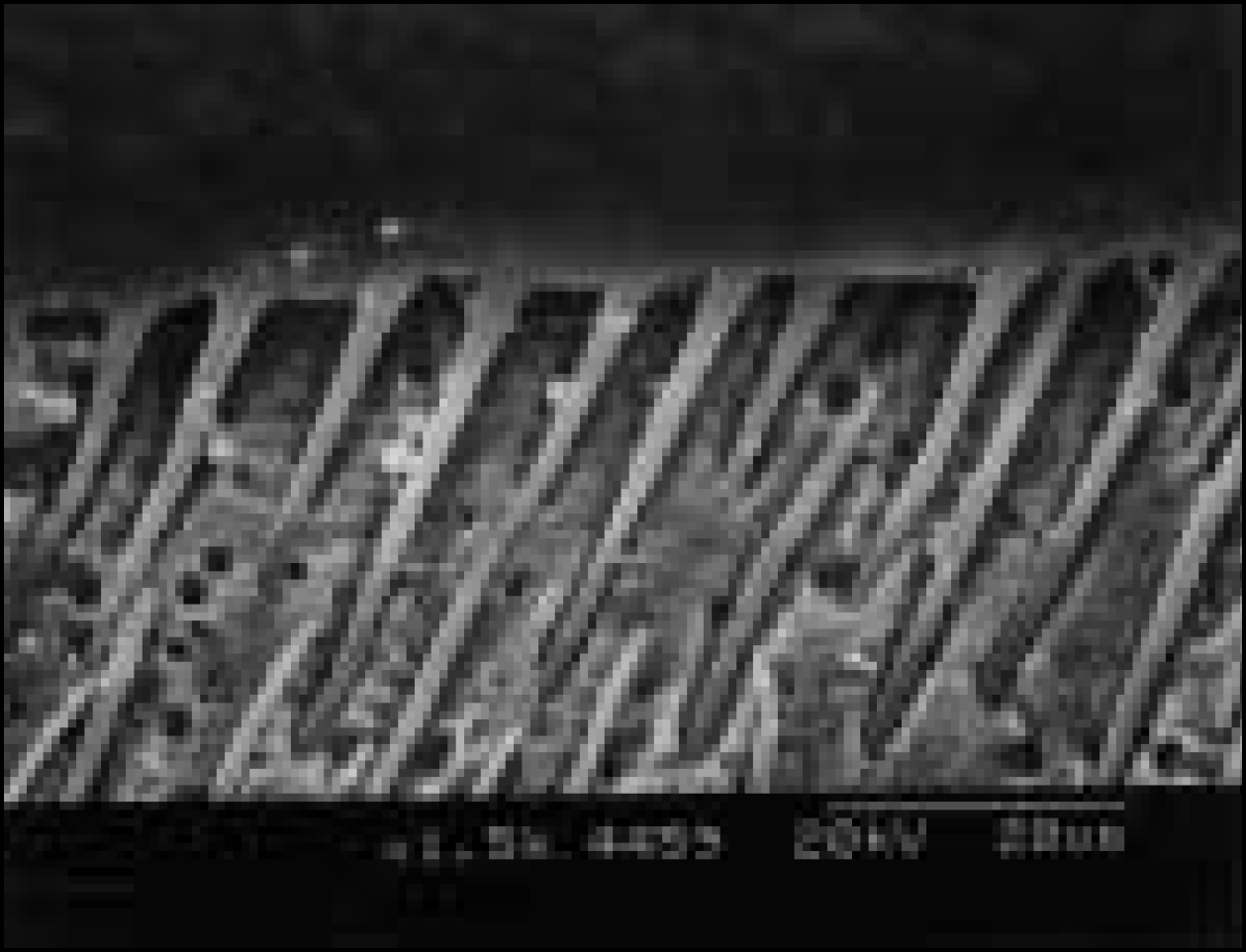
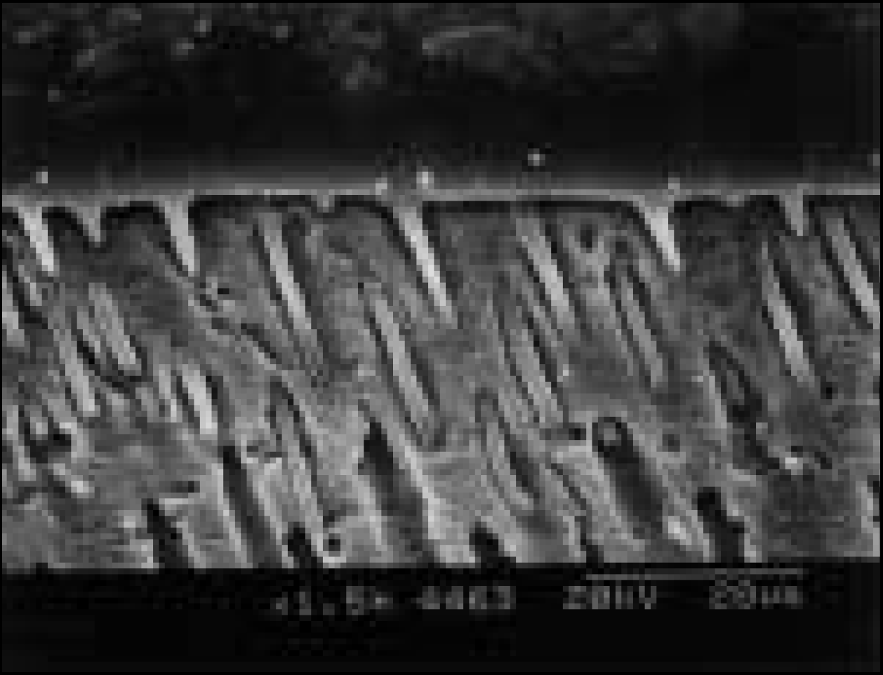
Table 1.
Materials used in this study
Table 2.
Experimental groups classified by luting methods, temporary cement, and dentin bonding system
Table 3.
Application manner of Dentin Bonding System (DBS)
Table 4.
Micro-shear bond strength of experimental group (Unit: ㎫, Mean ± S.D.)
 XML Download