Journal List > J Korean Acad Conserv Dent > v.30(3) > 1056158

Shin, Park, and Choi: Interleukin-8 and MCP(Monocyte Chemoattractant Protein)-1 expression by the Human Dental Pulps in cultures stimulated with Substance P

Abstract

The induction of the IL-8 and MCP-1 by the stimulation of Substance P and TNF-α (IL-8 agonist) and the specificity for SP using Spantide (SP antagonist) in the dental pulp tissues was measured quantitatively. In addition, the secretion of the IL-8 in the human dental pulp tissue 36 hrs after the stimulation of SP was observed after the stimulation of SP qualitatively.
According to this study, the results were as follows:
1. There was the significant IL-8 induction at 36 h after SP (10-4M) stimulation of the pulp tissue comparing with the unstimulated dental pulp tissues (p < 0.05). IL-8 immunostaining was weakly detected along the periphery of the pulp tissue after Mock stimulation and IL-8 immunostaining was detected around the fibroblast in the pulp tissue 36h After SP (10-4M) stimulation,
2. The secretion of MCP-1 from the dental pulp tissues comparing with Mock stimulation was induced at 36 hrs after TNF-α (40 ng/ml) stimulation, but no induction with SP(10-4M). TNF-α (40 ng/ml) did not induce the IL-8 secretion from the pulp tissue, weak IL-8 immunostaining was detected along the periphery of the pulp tissue.
3. Spantide (10-5M) inhibited IL-8 induction from the pulp tissues 36 h after SP (10-4M) stimulation.
These results suggest that SP significantly induces IL-8 recruiting neutrophils in localized human dental pulp tissue. MCP-1 appears to be less involved in the early establishment of pulpal inflammation in response to irritation such as mechanical insult of dentin. SP may have positive relation with the inflammation of the human dental pulp tissues.

I. INTRODUCTION

The causes of endodontic and pericpical pathosis are mechnical or chemical irritants and microbial infection of the pulp during dental procedures. A common pulpal response to the injuries is characterized by the influx of leukocytes to the affected sites1,2). The types of inflammatory pulpal conditions that are seen in response to external stimuli at cellular levels have been well described in earlier studies3).
Neutrophils are the most predominant cell population that migrates into inflammatory lesions. Neutrophil recruitment depends on chemotactic agonists that are synthesized and released at the site of inflammation; e.g., in a periodontal lesion. Chemotactic agonists may be derived from the host or from infecting microorganisms.
Chemokines are recently described family of chemotactic cytokines and reviewed by Graves and Jiang4). They are secondary pro-inflammatory mediators that are induced by primary pro-inflammatory mediators such as Interleukin-1 (IL-1) or tumor necrosis factor (TNF).
There are two families of chemokines based on the first cysteine residues. The α-chemokines, also known as CXC chemokines(eg, Interleukin-8), contain a single amino acid between the first and second cysteine resideues and are all located on chromosome 4. β, or CC chemokines have adjacent cysteine residues and located on chromosome 17. Most CXC chemokines are chemoattractants for neutrophils whereas CC chemokines generally attract monocytes, lymphocytes, basophils, and eosinophils.
Unlike the classic leukocyte chemoattractants, they exhibit a relatively high degree of specificity. Mononuclear phagocytes are thought to be one of the predominant cellular source of both interleukin-8 (IL-8) and monocyte chemoattractant protein-1 (MCP-1). However, IL-8 and MCP-1 can also be produced by nonimmune cells, such as fibroblast, keratinocytes, and endothelial cells, in response to both endogenous and exogenous stimuli5).
In addition, cytokines produced by mononuclear cells are important mediators for the cells (eg, dental pulp fibroblasts) to produce chemokine in the presence of microorganism6).
Interleukin-8 (IL-8) is the most thoroughly characterized neutrophil-stimulating chemokine and displays a wide range of biological effects, including chemotaxis and activation of neutrophils and it was originally described as a 72 amino acid peptide produced by human peripheral blood monocytes stimulated in culture with E.coli lipopolysaccharide7). The peripheral blood mononuclear cells were stimulated with microbes to evaluate the induction of a dose-dependent increase in chemokines8-9).
Expression of MCP-1 has been found in bacterial infections, specifically in gingival inflammation10-11) and osseous inflammation associated with lesions of endodontic origin12).
MCP-1 stimulates chemotaxis of monocytes, but on the other hand it does not stimulate chemotaxis of neutrophils. Local production of chemoattractants induce recruitment of leukocytes from the peripheral vasculature and leukocyte adhesion on, and migration along interstitial tissue in response to a chemotactic gradient.
The roles of Neurogenic inflammation in pulpal inflammation were reviewed in the past studies13-15). The richly innervated dental pulp expresses neuropeptides, the release of which (e.g., following injury) causes increased blood flow, plasma extravasation, and leukocyte accumulation leading to local inflammation. Leukocyte migration across the blood vessel wall into local tissue is a highly regulated process16). Local release of chemokines interleukin-8 (IL-8), a potent neutrophil chemoattractant, and monocyte chemoattractant protein-1 (MCP-1) plays a key role in attracting leukocytes to the sites of tissue injury.
Substance P (SP) and calcitonin gene-related peptide (CGRP) are two important sensory neuropeptides expressed in the dental pulp17). Although the primary function of these two neuopeptides, upon release, is to induce vasodilatation and pulpal blood flow17), they may play a more direct role in initiating the local inflammatory cell infiltration18). Substantial evidence has shown that neuropeptides can stimulate the production of proinflammatory cytokines by epithelial cells or fibroblasts from different tissues. SP or CGRP induces corneal epithelial cells to secrete IL-820,21) and bronchial epithelial cells to synthesize and release IL-6, IL-8 and TNF-α21). In addition, SP induces IL-8 production by fibroblasts in patients with osteoarthritis, while CGRP increases IL-8 and IL-6 secretion from fibroblasts in patients with rheumatoid arthritis22).
Recent study reported whether the cultured human pulp cells increase IL-8 secretion in response to SP stimulation23). In the present study, whether induction of IL-8 or MCP-1 in pulp tissue can be detected using enzyme-linked immunosorbent assay (ELISA) with ex vivo pulpal explants exposed to neuropeptides in culture and the IL-8 expression using immunohistochemical analysis with the ex vivo pulpal explants exposed to neuropeptides was evaluated.
To investigate further mechanisms that may contribute to leukocyte recruitment in lesions of endodontic origin, the differential expression of IL-8 and MCP-1 by human dental pulp tissues stimulated in vitro by the Substance P was examined.

II. MATERIALS AND METHODS

Sample preparation

Freshly extracted, intact, caries-free third molars were obtained from the patients (15~25 years old) in the Department of Oromaxillofacial Surgery, Kyung Hee medical center. Immediately after extraction, teeth were stored in phosphate buffered saline (PBS) and transferred to the laboratory. Under water coolant, each tooth was grooved longitudinally with a fissure bur at high speed. The tooth was then split with the driver, and the entire pulp(coronal and radicular) elevated with cotton pliers, maximum pulpal tissue was obtained. Some samples were divided into several small fragments approximately 2 × 2 mm in size each.
Repeated washing with PBS, pulp fragments were placed in a 60 mm culture dish containing Dulbecco's Modified Eagle Medium (DMEM; Life Technologies/GIBCO BRL, Gaithersburg, MD) supplemented with 10% fetal bovine serum (FBS). The collected pulp tissues were stored in DMEM at least 24 hrs before the stimulation with the Substance P. Cell culture media were supplemented with 100 units/ml penicillin-G, 100 µg/ml streptomycin, and 0.25 µg/ml fungizone (Gemini Bio-Products, Inc., Woodland, CA).
Some samples were divided in half, the 2 halves being stored separately under different conditions according to the analysis used (ELISA or immunocytochemistry). Samples for the ELISA were placed in a microcentrifuge tube containing 0.2 ml of DMEM without FBS and frozen at -70℃ until the assay. Samples for the immunohistochemical analysis were immediately either embedded in O.C.T. compound (Tissue-Tek, Miles Laboratories, Elkart, Ind), snap-frozen in liquid nitrogen, and stored at -70℃ until the preparation of frozen sections or placed in 10% phosphate-buffered formalin.

Stimulation of pulp tissues with Substance P

Substance P (10-4 M) or TNF-α(40 ng/ml) or 0.1% BSA (Mock stimulation) were added to the pulp tissue samples. Because of the small amount of sample volume (pulpal tissue in 200 µl of medium) and volume loss during the homogenization procedure, the supernatant of each sample was tested only once. Each test required 100 µl for each of 3 different dilution for IL-8 and MCP-1 ELISA test. The weight of the pulp tissue was measured before stimulation, and incubated at 37℃ in a humidified atmosphere of 5% CO2 and 95% air for 36 h. The pulp tissue including medium was collected and transferred to Micro Centrifuge tube. Pulpal samples were homogenized through use of Micro Centrifuge Sample Pestle (Scienceware, distributed by Fisher Scientific, Pittsburgh, PA). The dispersed tissue was centrifuged at 10,000 g for 5 min, weight measured and the supernatant collected. The amount of IL-8 and MCP-1 present in the supernatant was determined by means of ELISA.

Enzyme Linked Immunosorbent Assay for IL-8 and MCP-1

Standard ELISA for IL-8 was performed as described previously22) using 2 g/ml of polyclonal goat anti-human IL-8 (R&D Systems, Minneapolis, MN) as capturing antibodies, 1 g/ml polyclonal rabbit anti-human IL-8 (Endogen Inc., Cambridge, MA) as detecting antibodies, and 0.1 g/ml horseradish peroxidase (HRPO)-labeled polyclonal goat anti-rabbit immunoglobulin G (Biosource International, Camarillo, CA) as a secondary antibody. Subsequently fresh developing buffer containing substrate of optimal concentrations of TMB (3,3',5,5'-Tetramethyl benzidine ; Sigma, St. Louis, MO), H2O2 and sodium acetate, pH 6.0 was added and the developing reaction was stopped with 1.2 mol/L sulfuric acid. Absorbance at 450 nm was determined with a microplate reader (Bio-Tek Instrument, Inc., Laguna Hills, CA) and the concentrations were derived using the Delta Soft III software (Bio-Tek Instrument, Inc.). Known concentrations of recombinant human (rh) IL-8 (Endogen Inc., Cambridge, MA) was used to establish a standard curve for determining the concentrations of the experimental samples.
ELISA for MCP-1 was similar to that for IL-8. The following antibodies were used: monoclonal mouse anti-human MCP-1 (R&D Systems) as capturing antibodies, polyclonal rabbit anti-human MCP-1 (Cell Sciences Inc., Norwood, MA) as detecting antibodies, and HRPO-labeled polyclonal goat anti-rabbit immunoglobulin G (Biosource International, Camarillo, CA) as a secondary antibody. Known concentrations of rh-MCP-1 (R&D Systems) was used to establish a standard curve.

Immunohistochemistry

After the samples were fixed in 10% phosphate-buffered formalin for less than 2 weeks and then soaked in 30% sucrose in the microcentrifuge tube to inhibit the crytallization of the sample until the samples were precipitated at the bottom. The samples for the immunohistochemical analysis were immediately either embedded in snap-frozen O.C.T. compound (Tissue-Tek, Miles Laboratories, Elkart, Ind) at -20℃. Cryostat section (10 µm thick) were mounted on Vectabond precoated slides (Vector Laboratories, Inc, Burlingame, Calif). Sample sections that contained the greatest tissue area were prepared for staining with hematoxylin and eosin in addition to immunihistochemical staining. The sections were air dried for 12-24 hours before staining.
Immunoperoxidase staining was performed through use of the HRP-Envision system (Dako Corp, Carpinteria, CA). The sections were then washed 3 times (for 5 minutes each time) with wash buffer (Dako Corp, Carpinteria, CA) containg 50 mM Tris-HCl, 150 mM NaCl, 0.05% Tween 20 and a preservative, pH 7.6, and incubated in 3% H2O2 at room temperature for 5 minutes to block endogenous peroxidase. The sections were then washed 3 times (for 5 minutes each time) with wash buffer and then incubated for 30 minutes in a 0.1 % bovine serum albumin (BSA) of wash buffer. The sections were then washed 3 times with wash buffer and incubated at room temperature for 30 minutes with specific rabbit anti-human IL-8 polyclonal antibody (Endogen, Inc) that had been diluted 1 : 20 in wash buffer. Negative control experiments were performed for each sample by replacing the primary antibody with normal nonimmune goat serum at room temperature for the same time with the primary antibody. After the section were washed, the sections were again incubated with Peroxidase labeled polymer, HRP at room temperature for 30 minutes.
Following the wash, the site of peroxidase activity were visualized by incubation in 3,3'-Diaminobenzidine (DAB) solution for 10 minutes. After the final wash, sections were conterstained with hematoxyline, dipped 10 times into a bath of 37 mM ammonia water, and washed in dH2O for 5 minutes and then covered with a glass coverslip with a aqueous mounting solution (Dako Corp, Carpinteria, CA).

III. RESULTS

Enzyme Linked Immunosorbent Assay for IL-8 and MCP-1

Based on the preliminary studies showing that SP (10-4 M) increased minimal or no IL-8 secretion after 12 h, human dental pulp fragments were incubated for 36 h and 48 h after adding SP (10-4 M) or 0.1% BSA (Mock stimulation) or TNF-α (40 ng/ml) and a high concentration (10-4 M) of SP was used to maximize the induction. Increased IL-8 production was detected in pulp explants 36 and 48 hours after SP stimulation as shown in Figure 1. There were significant difference of IL-8 secretion between the Mock stimulation and SP stimulated groups after 36 hours (p < 0.05) (Figure 2), whereas no significant difference between two groups after 48 hours (p > 0.05) due to great variations of the base line levels of IL-8 from the Mock stimulated pulp fragments (either within the same tooth or between teeth) (Figure 3). However, the majority of the pulp fragments (5 out of 6) showed an increase of IL-8 (up to 5-fold) after SP stimulation (Figure 1). The specificity of this IL-8 induction in pulp explants by SP was verified by Spantide I (10-5 M), the presence of which inhibited IL-8 secretion which is same level with base-line (Mock stimulation), but there were no statistical significancy (P > 0.05). It was surprised that TNF-α induced no secretion of IL-8 from dental pulp tissues (Figure 1 and 4).
After the IL-8 ELISA from the dental pulp, the samples of the supernatant were used to determine whether an increase of MCP-1 could be detected in pulp explants exposed to SP. In contrast to the IL-8 induction in pulp explants as presented in Figure 1 and 2, MCP-1 increase was only detected in 50% (3 out of 6) pulp explant samples after 48 h of SP (10-4 M) stimulation (data was not shown), and the mean values show no induction (Figure 5). SP (10-4 M), Spantide (10-5 M) and Mock stimulation did not induce any MCP-1 secretion, but there was 1.8-fold increase of MCP-1 secretion at the stimulation of TNF-α (40 ng/ml) from the human dental pulp fragments (data was not significantly different, t-test, p > 0.05) (Figure 6).

Immunohistochemistry for IL-8

Experiments utilizing a monospecific IL-8 antiserum were carried out to determine whether the explant pulp tissue express IL-8 induction after SP stimulation for 36 hrs immunohistochemically. Out of the ELISA data, the secretion of MCP-1 was not induced by the stimulation of SP, which is the reason why MCP-1 immunohistochemistry was not examined. Because of the consistency in the secretion of the IL-8 from the pulp explants after 36 hrs comparing with 48 hrs, only 36 hrs samples were used in this experiments.
Frozen sections from the explant pulp tissues after the stimulation of Mock and SP (10-4 M), and TNF-α(40 ng/ml) were examined by incubation with IL-8 antiserum.
The normal pulp tissue (Mock stimulation) were incubated with the non-immunized control normal goat serum 36 hrs after the stimuation with DMEM (0.1% BSA) and negative IL-8 staining is displayed in the central pulp zone (Figure 7 A). To determine whether IL-8 positive cells were present in the normal pulp tissue without any stimulation (Figure 7 B), immunohistochemistry was carried out using a specific antiserum to IL-8. The outer cell-rich layer demonstrates weak IL-8 immunoreactivity (brown-colored precipitates) along the periphery of the pulp tissues. These immunohistologic findings on IL-8 staining in normal pulps may explain why in ELISA studies IL-8 was detected in normal samples (Figure 1).
In the pulp tissues stimulated with Substance P (10-4 M) for 36 hrs, negative IL-8 staining of the pulp tissues was seen in the central pulp zone (Figure 8 A). IL-8 staining of the pulp tissues were positive and IL-8 secretion from the fibroblasts in the pulp tissue were predominant. The intensity of IL-8 staining of the pulp tissues was varied from weak to marked (Figure 8 B).
In the pulp tissue incubated with TNF-α(40 ng/ml) for 36 hrs, there was no IL-8 staining of the tissue similar with the Mock stimulation, and no differences between the negative control (Figure 9 A) and IL-8 staining (Figure 9 B).

IV. DISCUSSION

The present study sheds light on the role of neuropeptides SP and CGRP in regulating the expression of the chemokines IL-8 and MCP-1 in dental pulp tissue. Although CGRP is highly expressed in the human dental pulp, it does not appear to play a significant role in inducing the expression of IL-8 and MCP-1 in dental pulp. While SP at the physiological level (10-10 to 10-8 M) does not appear to induce MCP-124-26), it shows a strong potential to induce IL-8 in human dental pulps. The data support the possibility that IL-8 induced by SP plays a more important role than MCP-1 in the early response of dental pulp to irritations by establishing local inflammatory cell infiltration.
It was previously found that IL-8 induction in cultured pulp cells can be observed at concentrations between 10-12 to 10-4 M of SP stimulation. This present studies further demonstrated that IL-8 levels in pulp tissue explants increased following SP stimulation. The induction of IL-8 observed in pulp explants required a longer time (36 h) to occur (Figure 2) in contrast to induction in cultured pulp cells (within 4 h). It is possible that the diffusion of SP into the pulp tissue required more time and therefore, it took longer to observe the increased IL-8 level in the pulp. The variation of IL-8 levels among pulp tissues from different teeth, as well as within the same tooth, makes the difference of IL-8 level between the Mock- and SP-stimulated groups difficult to measure. Nevertheless, mean values were consistently higher in the SP-stimulated groups. Although SP appears to be a potent inducer of IL-8 in pulp tissue, it requires a high dose (10-4 M) of amount to induce a mild MCP-1 induction. In comparison, CGRP at a high concentration of 10-4 M induced negligible IL-8, while inducing a moderate MCP-1 production in pulp cells.
Taken together, considering the physiological levels SP and CGRP are found in the human dental pulp (10-10 to 10-8 M)25,26), CGRP is not likely to play a significant role in initiating the inflammatory cell infiltration in dental pulp through inducing IL-8 or MCP-1. However, CGRP may participate in the process by inducing chemotactic response of T cells or dendritic cells27,28). In contrast, SP plays an important role in neurogenic inflammation due to its direct chemotaxis effect on neutrophils29) along with its potential to induce IL-8 production in pulp.
Local accumulation of neutrophils in the pulp underneath the invading caries or the dentin cavity filled with bacterial factors has been clearly demonstrated2). Bacterial components can serve as a potent chemoattractant to neutrophils. However, in a situation where bacteria may not be present and the dentin is only damaged mechanically, such as cavity or crown preparation, neutrophils egress from blood vessels and accumulation in the local pulp tissue underlying the damaged dentin can also be observed30). SP receptor neurokinin-1 (NK1) is expressed in cells of the cell-rich zone beneath the odontoblast layer31), suggesting that when SP is released from the excited sensory fibers, these cells may be activated by SP to produce IL-8.
It seems logical to link SP, IL-8 and neutrophils as three important early responsive players following imposition of the external stimuli on the pulpodentin complex. The fast-responding neural system releases SP rapidly upon stimulation inducing local tissue cells to produce and secrete IL-8, which in turn attracts neutrophils to migrate toward the irritated sites. This sequence of events leads to the formation of neutrophil infiltration in the microenvironments localized acute inflammatory response. In contrast to the role of IL-8 which preferentially attracts and stimulates neutrophils, MCP-1 attracts and stimulates mainly monocytes4). Therefore, our findings of no significant MCP-1 induction by either neuropeptides appear to fit into the scheme of the early stage of inflammation in the pulp that more neutrophils, than macrophages, are recruited into the localized area subjacent to the irritated dentin.

V. CONCLUSION

The purpose of the present study was to examine the coordinate activity between neuropeptide and cytokine, and their important role in sensing and eliciting rapid immune response to the external irritations to the dental pulp. For the purpose, the induction of the IL-8 and MCP-1 by the stimulation of Substance P and TNF-α (IL-8 agonist) and the specificity for SP using Spantide (SP antagonist) in the dental pulp tissues was measured quantitatively. In addition, the secretion of the IL-8 in the human dental pulp tissue 36 hrs after the stimulation of SP was observed after the stimulation of SP qualitatively.

According to this study, the results were as follows:

  1. There was the significant IL-8 induction at 36 h after SP(10-4 M) stimulation of the pulp tissue comparing with the unstimulated dental pulp tissues(p < 0.05).

  2. The secretion of MCP-1 from the dental pulp tissues comparing with Mock stimulation was induced at 36 hrs after TNF-α (40 ng/ml) stimulation, but no induction with SP (10-4 M).

  3. Spantide (10-5 M) inhibited IL-8 induction from the pulp tissues 36 h after SP (10-4 M) stimulation.

  4. IL-8 immunostaining was weakly detected along the periphery of the pulp tissue 36 h after Mock stimulation.

  5. 36h After SP (10-4 M) stimulation, IL-8 immunostaining was detected around the fibroblast in the pulp tissue.

  6. TNF-α (40 ng/ml) did not induce the IL-8 secretion from the pulp tissue, weak IL-8 immunostaining was detected along the periphery of the pulp tissue.

These results suggest that SP significantly induces IL-8 expression in human dental pulp suggesting an important role of SP in recruiting neutrophils in localized pulp tissue. MCP-1 appears to be less involved in the early establishment of pulpal inflammation in response to irritation such as mechanical insult of dentin. SP may have positive relation with the inflammation of the human dental pulp tissues.

Figures and Tables

Figure 1
IL-8 secretion after stimulation of the pulp tissues with Substance P (10-4 M) and TNF-α(40 ng/ml) for 36 and 48 hrs. Data are mean ± SEM of six independent experiments of 1-6 sets of pulp fragments (each set was obtained from one tooth).
*Statistically significant, P < 0.05.
jkacd-30-193-g001
Figure 2
IL-8 secretion after stimulation of the pulp tissues with Substance P (10-4 M) for 36 and 48 hrs. Data are mean ± SEM of six independent experiments of 1-6 sets of pulp fragments(each set was obtained from one tooth).
jkacd-30-193-g002
Figure 3
IL-8 secretion after stimulation of the pulp tissues with Substance P (10-4 M) and Spantide (10-5 M) for 36 hrs. Data are mean ± SEM of six independent experiments of 1-6 sets of pulp fragments (each set was obtained from one tooth).
jkacd-30-193-g003
Figure 4
IL-8 secretion after stimulation of the pulp tissues with TNF-α(40 ng/ml) in the course of time. Data are mean ± SEM of six independent experiments of 6 pulp fragments.
jkacd-30-193-g004
Figure 5
MCP-1 secretion after stimulation of the pulp tissues with Substance P (10-4 M) and TNF-α(40 ng/ml) for 36 and 48 hrs. Data are mean ± SEM of six independent experiments of 1-6 sets of pulp fragments(each set was obtained from one tooth).
jkacd-30-193-g005
Figure 6
MCP-1 secretion after stimulation of the pulp tissues with TNF-α(40 ng/ml) in the course of time. Data are mean ± SEM of six independent experiments of 6 pulp fragments.
jkacd-30-193-g006
Figure 7
Expression of IL-8 in the pulp tissue after mock stimulation. (Original magnification, × 200)
A. Frozen sections were incubated with IL-8 antiserum.
B. Frozen sections were incubated with nonimmunized normal goat serum.
jkacd-30-193-g007
Figure 8
Expression of IL-8 in the pulp tissue after SP (10-4M) stimulation. (Original magnification, × 200)
A. Frozen sections were incubated with IL-8 antiserum.
B. Frozen sections were incubated with nonimmunized normal goat serum.
jkacd-30-193-g008
Figure 9
Expression of IL-8 in the pulp tissue after TNF (40 ng/ml) stimulation. (Original magnification, × 200)
A. Frozen sections were incubated with IL-8 antiserum.
B. Frozen sections were incubated with nonimmunized normal goat serum.
jkacd-30-193-g009

References

1. Stanley HR. Importance of the leukocyte to dental health. J Endod. 1977. 3:334–341.
2. Bergenholtz G, Lindhe J. Effect of soluble plaque factors on inflammatory reactions in the dental pulp. Scand J Dent Res. 1975. 83:153–158.
crossref
3. Seltzer S, Bender IB, Ziontz M. The dynamics of pulp inflammation: correlations between diagnostic data and actual histologic findings in the pulp. Oral Surg Oral Med Oral Pathol. 1963. 16:846–871.
crossref
4. Graves DT, Jiang Y. Chemokines, a family of chemotactic cytokines. Crit Rev Oral Biol Med. 1995. 6:109–118.
crossref
5. Rauschenberger C, Bailey J, Cootauco C. Detection of human IL-2 in normal and inflamed dental pulps. J Endod. 1997. 23:366–370.
crossref
6. Nagaoka S, Yokuda M, Sakuta T, Taketoshi Y, Tamura M, Kakada H, Kawagoe M. Interleukin-8 gene expression by human dental pulp fibroblast in cultures stimulated with Prevotella intermedia Lipopolysaccharide. J Endod. 1996. 22:9–12.
crossref
7. Baggiolini M, Walz A, Kunkel SL. Neutrophil-activating peptide-1/interleukin 8, a novel cytokine that activates neutrophils. J Clin Invest. 1989. 84:1045–1049.
crossref
8. Peveri P, Walz A, Dewald B, Baggiolini M. A novel neutrophil activating factor produced by human mononuclear phagocytes. J Exp Med. 1988. 167:1547–1559.
crossref
9. Jiang Y, Russel TR, Graves DT, Cheng H, Nong SH, Levitz SM. Monocyte chemoattractant protein 1 and Interleukin-8 production in mononuclear cells stimulated by oral microorganisms. Infect Immun. 1996. 64:4450–4455.
crossref
10. Yu X, Antoniades HN, Graves DT. Expression of monocyte chemoattractant protein 1 in human inflamed gingival tissues. Infect Immun. 1993. 61:4622–4628.
crossref
11. Tonetti MS, Imboden MA, Gerber L, Lang NP, Laissue J, Mueller C. Localized expression of mRNA for phagocyte-specific chemotactic cytokines in human periodontal infection. Infect Immun. 1994. 62:4005–4014.
crossref
12. Rahimi P, Wang CY, Stashenko P, Lee SK, Lorenzo JA, Graves DT. Monocyte Chemoattractant protein-1 expression and monocyte recruitment in osseous inflammation in the mouse. Endocrinology. 1995. 136:2752–2759.
crossref
13. Jontell M, Okiji T, Dahlgren U, Bergenholtz G. Immune defense mechanisms of the dental pulp. Crit Rev Oral Biol Med. 1998. 9:179–200.
crossref
14. Stashenko P, Teles R, D'Souza R. Periapical inflammatory responses and their modulation. Crit Rev Oral Biol Med. 1998. 9:498–521.
crossref
15. Byers MR, Narhi MV. Dental injury models; esperimental tools for understanding neuroinflammatory interactions and polymodal nociceptor functions. Crit Rev Oral Biol Med. 1999. 10:4–39.
crossref
16. Muller WA, Randolph GJ. Migration of leukocytes across endothelium and beyond: molecules involved in the transmigration and fate of monocytes. J Leukoc Biol. 1999. 66:698–704.
crossref
17. Wakisaka S. Neuropeptides in the dental pulp: distribution, origins, and correlation. J Endod. 1990. 16:67–69.
crossref
18. Fristad I, Kvinnsland IH, Jonsson R, Heyeraas KJ. Effect of intermittent long-lasting electrical tooth stimulation on pulpal blood flow and immunocompetent cells: a hemodynamic and immunohistochemical study in young rat molars. Exp Neurol. 1997. 146:230–239.
crossref
19. Tran MT, Lausch RN, Oakes JE. Substance P differentially stimulates IL-8 synthesis in human corneal epithelial cells. Invest Ophthalmol Vis Sci. 2000. 41:3871–3877.
20. Tran MT, Ritchie MH, Lausch RN, Oakes JE. Calcitonin gene-related peptide induces IL-8 synthesis in human corneal epithelial cells. J Immunol. 2000. 164:4307–4312.
crossref
21. Veronesi B, Carter JD, Devlin RB, Simon SA, Oortgiesen M. Neuropeptides and capsaicin stimulate the release of inlfammatory cytokines in a human broncheal epithelial cell line. Neuropeptides. 1999. 33:447–456.
crossref
22. Raap T, Justen HP, Miller LE, Cutolo M, Scholmerich J, Straub RH. Neurotransmitter modulation of interleukin 6 (IL-6) and IL-8 secretion of synovial fibroblasts in patients with rheumatoid arthritis compared to osteoarthritis. J Rheumatol. 2000. 27:2558–2565.
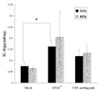
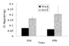
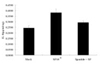
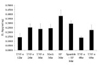
23. Patel T, Park SH, Lin LM, Ciappelli F, Huang GT. Substance P induces interleukin-8 secretion from human dental pulp cells. Oral Surg Oral Med Oral Pathol Oral Radiol Endod. 2003. 96:478–485.
crossref
24. Hargreaves KM, Swift JQ, Roszkowski MT, Bowles W, Gary MG, Jackson DL. Pharmacology of peripheral neuropeptide and inflammatory mediator release. Oral Surg Oral Med Oral Pathol. 1994. 78:503–510.
crossref
25. Awawdeh L, Lundy FT, Shaw C, Lamey PJ, Linden GJ, Kennedy JG. Quantitative analysis of substance P, neurokinin A and calcitonin gene-related peptide in pulp tissue from painful and healthy human teeth. Int Endod J. 2002. 35:30–36.
crossref
26. Bowles WR, Withrow J, Lepinski A, Hargreaves KM. Tissue levels of immunoreactive substance P are increased in patients with irreversible pulpitis. J Endod. 2003. 29:265–267.
crossref
27. Foster CA, Mandak B, Kromer E, Rot A. Calcitonin gene-related peptide is chemotactic for human T lymphocytes. Ann N Y Acad Sci. 1992. 657:397–404.
crossref
28. Dunzendorfer S, Kaser A, Meierhofer C, Tilg H, Wiedermann CJ. Cutting edge: peripheral neuripeptides attract immature and arrest mature blood-derived dendritic cells. J Immunol. 2001. 166:2167–2172.
crossref
29. Roch-Arveiller M, Regoli D, Chanaud B, Lenoir M, Muntaner O, Stralzko S, Giroud JP. Tachykinins: effects on motility and metabolism of rat polymorphonuclear leucocytes. Pharmacology. 1986. 33:266–273.
crossref
30. Takahashi K. Changes in the pulpal vasculature during inflammation. J Endod. 1990. 16:92–97.
crossref
31. Fristad I, Vandevska-Radunovic V, Kvinnsland IH. Neurokinin-1 receptor expression in the mature dental pulp of rats. Arch Oral Biol. 1999. 44:191–195.
crossref
TOOLS
Similar articles