

 PDF
PDF ePub
ePub Citation
 Print
Print


I. INTRODUCTION
Bacteria and their by-products are considered to be the primary etiologic agents of pulpal necrosis and periapical lesions1). Therefore, endodontic treatment aims at eliminating of microorganisms from the root canal system and preventing of reinfection. However some cases fail even when apparently well treated. In fact, a number of factors associated with failure of endodontic therapy. But, most treatment failures are caused by microorganisms persisting in the apical parts of root canals of obturated teeth. It seems to be bacterial effects in root canal and periapical lesion. Especially, single spices of gram-positive organisms, Enterococcus faecalis had been found to be one of predominant bacteria in teeth in which root canal therapy failed2).
So, the use of intracanal medication to disinfect the root canal system has been advocated3). Calcium hydroxide (Ca(OH)2) has been the intracanal medication of choice. Sjögren et al.4) and byström et al.3) reported that 7-days and 30-days applications of Ca(OH)2, respectively, eliminated bacteria that survived instrumentation in root canals. However, in case of E. faecalis, Ca(OH)2 dressing is ineffective3). Therefore it seems that the frequent isolation of Enterococci in failed endodontic cases puts in question the routine use of Ca(OH)2 as an intracanal medication5). Survival of E. faecalis in Ca(OH)2 at high pH was related to a functional proton pump7). However few researches reported how these factors affect the antibacterial activity of Ca(OH)2.
Matrix metalloproteinases (MMPs) form a family of structurally related but genetically distinct endopeptidases expressed at low level in normal tissue, but upregulated during inflammation8). They are group of zinc-dependent proteinases, secreted or released by host cells (ie, PMN, macrophage, bone cell, fibroblast) as proenzymes. There are multiple members of family, including interstitial collagenase (MMP-1), neutrophil collagenage (MMP-8), stromelysin-1 (MMP-3), stromelysin-2 (MMP-10), gelatinase B (MMP-9). Shin et al.9) reported that MMPs play an important role in the pulp tissue destruction in inflamed pulp and periapical lesion. MMP-8 of them has been thought to be uniquely produced by developing PMN cells in bone marrow, and that the MMP-8 activity is dependent on the release of the enzyme from PMN cells by degranulation10). However, recent findings demonstrated expression of MMP-8 in mesenchyme derived, non-PMN lineage cells, including dental pulp fibroblasts, odontoblasts, inflamed epithelial cells and plasma cells11-12). In other research, MMP-8 is reported that they have an important role in the degenerations of the extracellular matrix in inflamed pulps and periapical lesions13).
In this study, the purpose was to examine the levels of production of MMP-8 by PMN stimulated with Enterococcus faecalis and effect of Ca(OH)2 on their production.
II. MATERIALS AND METHOD
Preparation of bacterial sonicated extracts
Enterococcus faecalis (ATCC 29212) strains were used in this study. Strains were grown in 1-liter cultures in 85% N2-10% H2-5% CO2 chamber for 3 days at 37℃. The medium used was 3.7% brain heart infusion broth supplemented with 0.5% yeast extract, 0.5mg L-cysteine hydrochloride, and 0.5% sodium bicarbonate.
Bacterial fractions were prepared. Briefly, bacterial cell harvest from 1-liter cultures were washed and suspended in 20ml of phosphate-buffered saline (PBS). To minimize the loss of protein, 1mK of Phenylmethylsulfonylfluoride (PMSF) were added. PMSF act as protease inhibitor at protein extraction. And suspensions of bacterial cell were disrupted by sonication (100W output, Fisher sonicator) on ice for 5min with 30-sec pulses on and 10-sec pulse-off in the presence of glass beads. Sonicated debris was centrifuged at 12,000 × g in a Sorvall RC5C (Sorvall Instruments, DuPont) for 20 min, and membrane fraction was sedimented by ultracentrifugation at 85,000 × g for 60 min at 4℃. To investigate protein profile of sonicated extracts of E. faecalis (SEF), supernatant was filtered with 0.22 µmm syringe and separated by 10% SDS-PAGE at 100V for 90min, and the protein concentration was determined by Bicinchoninic acid (BCA) protein assay (Pierce, Rockford, IL). The SDS-PAGE reveals that the soluble cytoplasmic protein of E. faecalis strain ATCC 29212 is mainly consist of 75kDa and 35-50kDa polypeptide. The pellet was resuspended in 20 ml of 10mM Tris buffer (pH 7.4), and stocked in deepfreezer at -20℃.
Sonic extract of E. faecalis (SEF) treated with Ca(OH)2
Each of SEF was suspended to 100 µg/ml with pyrogen-free water. Ca(OH)2 powder (Sigma Chemical Co., St Louis, MO, USA) 12.5 mg/ml was added. All samples were vortexed for 20 seconds once a day and incubated at 37℃ for 7days. After that, upper part of solution was ultrafilterated (Centriprep YM-10, 10,000 NMWL, Millipore Co., USA) and stocked in deep-freezer at -20℃.
Preparation of PMN
Using heparinized tubes, Venous blood was obtained from 6 healthy volunteers (26~35years). The erythrocytes of the obtained blood were segregated through 6% dextran (Mwt. 500,000, Sigma Chemical Co., St Louis, MO, USA), the leukocytes were extracted after centrifugation using lymphoprep (Nycomed Pharma AS, Oslo, Norway) and remained erythrocytes were eliminated by carrying out hypotonic lysis. After verification through the Trypan blue dye exclusion test & Giemsa staining, the PMN leukocytes that were obtained by this method proved to have 98% or more vitality and 97% or more purity. One millimeter of the extracted PMN leukocytes were 24 well plate with each well having a cell account of 10 × 104.
Bacterial stimulation and MMP-8 production
SEF group and SEF treated with Ca(OH)2 group and E.coli LPS were diluted at three concentration. And PMN media were stimulated by each groups and control group was not activated. Final concentrations of each group were 0.05 µg/ml, 0.5 µg/ml and 5 µg/ml and incubated at 37℃ for 30 minutes.
III. RESULTS
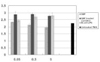
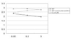
The results of this study are the mean values of six samples of duplicate cultures and summarized in Table 1, Figure 1 and Figure 2.
In the SEF groups, the level of production of MMP-8 was higher in comparison to the negative control group in low concentration (0.05 µg/ml) of SEF (P < 0.05), but it decreased with an increase in the concentration of SEF (P < 0.05). In the case of SEF treated with Ca(OH)2, all of the MMP levels at different SEF concentrations were higher than control group (P < 0.05), but no statistical difference was found among the different SEF concentrations (P > 0.05). All of the levels in E. coli LPS were increased with increasing concentrations (P < 0.05).
IV. DISCUSSION
In recent years, MMPs have gained considerable attention in many studies. MMPs play an important role in the pulp tissue destruction of acute inflamed pulp9). The role of MMP-8 synthesized by the cells in the pulp-dentinal complex is not clear. But MMP-8 was reported that they have an important role in the degenerations of the extracellular matrix in inflamed pulps and periapical lesions13). Therefore, a hypothesis was proposed that MMP-8 could be present in the periapical lesion, and the enzyme level could be related to the activity of the periapical tissue. And MMPs are endopeptidases expressed at low level in normal tissues, but upregulated during inflammation8). So, untreated PMN had some MMP-8 in this study.
In previous studies, sonic extracts from bacteria were studied for their effects on the immune response. For example, sonicated material of Fusobacterium nucleatum can evoke a concentration-dependent stimulatory or suppressive effect on the proliferation rate of accessory cell14). Sonicated extract of Actinobacillus actinomycetemcomitans suppressed interleukin-2 production of T-cells15). Kim et al.16) showed that sonicated extracts of Enterococcus faecalis also suppressed the production of interleukin-2 and interleukin-4 in lymphocytes. So in this study, SBE may have depressive effect to PMN. In positive control group, all of the MMP-8 levels in E. coli LPS were increased with increasing concentrations (P < 0.05).
In this study, MMP-8 production level at low level of SEF (0.05µg/ml) was seen a little elevation compare to untreated control group (P < 0.05). But it decreased with an increase in the concentration of SEF (P < 0.05). This phenomenon showed that ability of E. faecalis to impair the production of MMP-8 may be related to the suppression of PMN. This fact may be seen another immunosuppressive effect of SEF.
However, there is no study how E. faecalis have immunosuppressive effect exactly. But E. faecalis is the most prevalent species of enterococci in root canals of failed case17) and for it to be involved in the pathogenesis and maintenance of apical periodontitis, it must survive in the root filled canals as where the nutrient supply is limited. E. faecalis has been shown to have relatively uncommon capacity to survive in root canals as a monoculture without the support of other bacteria18). E. faecalis is also known to survive periods of starvation in water19) and infected dentin in water for periods of at least 10 days20).
Ca(OH)2 has been widely used as an intracanal medication to eliminate bacteria and is currently acknowledged to be one of the most effective antimicrobial dressings used during endodontic therapy21). Although the exact mechanism of action of Ca(OH)2 is still unknown, its antimicrobial activity is generally considered to be related to the release of hydroxylions in an aqueous environment, producing a pH of approximately 12.5, even in very dilute mixture. Most endodontic pathogens are unable to survive in this alkaline environment21). The mechanism that Ca(OH)2 uses to eliminate bacteria may include damage to bacteria cytoplasmic membrane by including lipid peroxidation, protein denaturation, and damage to bacterial DNA and by serving as physical barrier that withholds nutrients for bacterial growth and limits space for bacterial multiplication22).
But E. faecalis is known to withstand a high pH. This is an identifying characteristic of E. faecalis. At pH 11.4 or greater, E. faecalis does not survive, yet it can survive at a pH below 11.5 as shown in this and earlier studies3). Because of the buffering effect of dentin23-25), it is unlikely that the high pH of Ca(OH)2 (> 11.5) is attained within dentinal tubules where E. faecalis has the capacity, at least in vitro, to penetrate deeply20,26-28). In radicular dentin, alkalinity may only reach pH 10.3 after dressing the canal with Ca(OH)224). An adaptive response in alkaline pH and stress-induced protein synthesis appear to play minor roles in cell survival, a functional proton pump with the capacity to acidify the cytoplasm is critical for survival of E. faecalis at high pH7).
So considered E. faecalis in endodontic treatment, additive medication is recommended. For example, chlorhexidine (CHX) is a broad-spectrum antimicrobial agent that has been reported to be an effective medicament in endodontic treatment therapy29). As a root canal irrigant and intracanal medicament, CHX has an antibacterial efficacy comparable to that of sodium hypochlorite (NaOCl)30-31). In addition, it is also effective against strains resistant to Ca(OH)232). Therefore the use of combination of Ca(OH)2 and CHX is recommended in endodontic treatment.
In our study, Ca(OH)2 had some effect to production of MMP-8 by PMN. But comparison to untreated PMN group, this data does not support that Ca(OH)2 removed immunosuppressive effect of SEF. MMP-8 levels of Ca(OH)2 treated SEF group had another impression, this may because of the high pH of Ca(OH)2. Gram positive microorganisms have external membrane structure, cell wall. This cell wall have two important high molecule, peptidoglycan and teicoic acid. In this study, SEF treated with Ca(OH)2 for 1 week showed different appearance in the production of MMP-8. This result means that Ca(OH)2 could possibly modified SEF. The high pH of Ca(OH)2 may modify acidity of teicoic acid. But no statistical difference was found among the different concentrations (P > 0.05).
In our study, this result suggested that bacterial protein sonicated extract of E. faecalis can inhibit the immune response by PMN and this could be one of possible factors why E. faecalis are found frequently in the teeth with failed endodontic treatment. The use of an antimicrobial agent as intracanal medication between appointments may increase the chances for successful endodontic treatment by reducing residual bacteria in root canal system. In endodontic treatment, Ca(OH)2 may have limited antibacterial effect to E. faecalis. So our study suggests that the use of only Ca(OH)2 in clinical endodontic treatment may not be effective to E. faecalis.
V. CONCLUSION
According to this study we could summarize as follows:
MMP-8 was expressed at low level in untreated PMN group and the levels of MMP-8 were upregulated in PMN stimulated by E. coli LPS groups.
In the SEF groups, the level of production of MMP-8 decreased with an increase in the concentration of SEF (p < 0.05). So E. faecalis may have suppressive effect on the production of MMP-8 by PMN.
In the case of SEF treated with Ca(OH)2, all of the MMP levels at different SEF concentrations were higher than SEF & untreated PMN groups (p < 0.05), but no statistical difference was found among the different SEF concentrations (p > 0.05).

 XML Download