

 PDF
PDF ePub
ePub Citation
 Print
Print


Abstract
This study was done to evaluate whether there were any differences in microleakage of class V composite restorations according to restoration site and cavity size.
Total sixty-four restorations were made in molar teeth using Esthet-X. Small (2 × 2 × 1.5 mm) and large (4 × 2 × 1.5 mm) restorations were made at the buccal/lingual surface and the proximal surface each. After 1,000 times of thermocycling (5℃ - 55℃), resin replica was made and the percentage of marginal gap to the whole periphery of the restoration was estimated from SEM evaluation.
Thermocycled tooth was dye penetrated with 50% silver nitrate solution. After imbedding in an auto-curing resin, it was serially ground with a thickness of 0.25 mm. Volumetric microleakage was estimated after reconstructing three dimensionally.
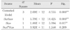
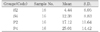
Two-way ANOVA and independent T-test for dye volume, Mann-Whitney U test for the percentage of marginal gap, Spearman's rho test for the relationship between two techniques were used.
The results were as follows:
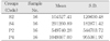
1. The site and size of the restoration affected on the microleakage of restoration. Namely, much more leakage was seen in the proximal and the large restorations rather than the buccal/lingual and the small restorations.
2. Close relationship was found between two techniques (Correlation coefficient = 0.614 / P = 0.000).
Within the limits of this study, it was noted that proximal and the large restorations leaked more than buccal/lingual and the small restorations. Therefore, it should be strictly recommended large exposure of margins should be avoided by reducing unnecessary tooth reduction.
Figures and Tables
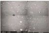 | Figure 2Calculation of marginal gap: Percentage of marginal gap to cavity perimeter (SEM image ; × 35)
(Blue line : cavity perimeter, Red line : area with marginal gap)
|
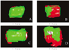 | Figure 3Internal view of three-dimensionally reconstructed images of experimental groups
(Green : restoration, Red : dye)
|


References
1. Eick JD, Welch FH. Polymerization shrinkage of posterior composite resins and its possible influence on postoperative sensitivity. Quintessence Int. 1986. 17(2):103–111.
2. Tjan AH, Bergh BH, Lidner C. Effect of various incremental techniques on the marginal adaptation of Class II composite resin restorations. J Prosthet Dent. 1992. 67(1):62–66.

3. Lutz F, Krejci , Oldenburg TR. Elimination of polymerization stresses at the margins of posterior composite resin restorations: A new restorative technique. Quintessence Int. 1986. 17(12):777–784.
4. Uno S, Asmussen E. Marginal adaptation of a restorative resin polymerized at reduced rate. Scand J Dent Res. 1991. 99:440–444.
5. Burgess JO, DeGoes M, Walker R, Ripps AH. An evaluation of four light-curing units comparing soft and hard curing. Pract Periodontics Aesthet Dent. 1999. 11(1):125–133.
6. Aboushala A, Kugel G, Hurley E. Class II composite resin restorations using glass-ionomer liners: Microleakage studies. J Clin Pediatr Dent. 1996. 21(1):67–71.
7. Õilo G. Biodegradation of dental composites/glassionomer cements. Adv Dent Res. 1992. 6:50–54.
8. Fortin D, Swift EJ Jr, Denehy GE, Reinhardt JW. Bond strength and microleakage of current dentine adhesives. Dent Mater. 1994. 10(4):253–258.
9. Rigsby DF, Retief DH, Bidez MW, Russell CM. Effect of axial load and temperature cycling on microleakage of resin restorations. Am J Dent. 1992. 5(3):155–159.
10. Donly KJ, Ellis RK. Glass inserts. A new dimension in restorative dentistry. Am J Dent. 1989. 2(1):21–24.
11. Feilzer A, de Gee AJ, Davidson CL. Setting stress in composite resin in relation to configuration of the restoration. J Dent Res. 1987. 66:1636–1639.
12. Hobson RS, Rugg-Gunn AJ, Booth TA. Acid-etch patterns on the buccal surface of human permanent teeth. Arch Oral Biol. 2002. 47(5):407–412.
13. Sturdevant JR, Pashley DH. Regional dentin permeability of Class I and II cavity preparations. J Dent Res. 1989. 68:203. (abstract No. 173).
14. Saboia Vde P, Pimenta LA, Ambrosano GM. Effect of collagen removal on microleakage of resin composite restorations. Oper Dent. 2002. 27(1):38–43.
15. Adamo HL, Buruiana R, Schertzer L, Boylan RJ. A comparison of MTA, Super-EBA, composite and amalgam as root end filling materials using a bacterial microleakage model. Int Endod J. 1999. 32:197–203.
16. Hembree JH, Andrew JT. Microleakage of several class V anterior restorative materials: a laboratory study. J Am Dent Assoc. 1978. 97(2):179–183.
17. Manhart J, Chen HY, Mehl A, Weber K, Hickel R. Marginal quality and microleakage of adhesive class V restorations. J Dent. 2001. 29(2):123–130.
18. Youngson CC, Jones JC, Fox K, Smith IS, Wood DJ, Gale M. A fluid filtration and clearing technique to assess microleakage associated with three dentine bonding systems. J Dent. 1999. 27(3):223–233.
19. Von Fraunhofer JA, Adachi EI, Barnes DM, Romberg E. The effect of tooth preparation on microleakage behavior. Oper Dent. 2000. 25(6):526–533.
20. Ha SY, Shin DH. New quantitative measuring technique for microleakage of the restored tooth through 3D reconstruction. J Korean Acad Conserv Dent. 2004. 29(5):413–422.
21. Estafan D, Pines MS, Erakin C, Fuerst PF. Microleakage of Class V restorations using two different compomer systems: an in vitro study. J Clin Dent. 1999. 10(4):124–126.
22. Hatibovic-Kofman S, Wright GZ, Braverman I. Microleakage of sealants after conventional, bur, and air-abrasion preparation of pits and fissures. Pediatr Dent. 1998. 20(3):173–176.
23. Grande RH, Ballester R, Singer Jda M, Santos JF. Microleakage of a universal adhesive used as a fissure sealant. Am J Dent. 1998. 11(3):109–113.
24. Mixson J, Eick JD, Chappell RP, Tira DE, Moore DL. Comparison of two-surface and multi-surface scoring methodologies for in vitro microleakage studies. Dent Mater. 1991. 7(3):191–196.
25. Shin DH. Dentinal microleakage study on the light curable restorative glass ionomer cement. J Korean Acad Conserv Dent. 1995. 20(2):832–838.
26. Arnold WH, Gaengler P, Kalkutschke L. Three-dimensional reconstruction of approximal subsurface caries lesions in deciduous molars. Clin Oral Investig. 1998. 2(4):174–179.
27. Mikrogeorgis G, Lyroudia KL, Nikopoulos N, Pitas I, Molyvdas I, Lambrianidis TH. 3D computer-aided reconstruction of six teeth with morphological abnormalities. Int Endod J. 1999. 32(2):88–93.
28. Jacobs R, Adriansens A, Verstreken K, Suetens P, van Steenberghe D. Predictability of a three-dimensional planning system for oral implant surgery. Dentomaxillofac Radiol. 1999. 28(2):105–111.
29. Jung EH, Shin DH. Morphological analysis of Cshaped root using 3D reconstruction. J Korean Acad Conserv Dent. 2002. 27(4):421–431.
30. Veis A, Lambrianides T, Nicolaou A. Area-metric analysis of dye leakage for evaluation of sealing ability of root canal obturation techniques. Endod Dent Traumatol. 1996. 12(5):222–226.
31. Gale MS, Darvell BW, Cheung GS. Three-dimensional reconstruction of microleakage pattern using a sequential grinding technique. J Dent. 1994. 22(6):370–375.
32. Dorminey JC, Dunn WJ, Taloumis LJ. Shear bond strength of orthodontic brackets bonded with a modified 1-step etchant-and-primer technique. Am J Orthod Dentofacial Orthop. 2003. 124(4):410–413.
33. Carvalho RM, Pereira JC, Yoshiyama M, Pashley DH. A review of polymerization contraction: The influence of stress development versus stress relief. Oper Dent. 1996. 21:17–24.
34. Asmussen E, Munksgaard EC. Bonding of restorative resins to dentine: Status of dentin adhesives and impact on cavity design and filling techniques. Int Dent J. 1988. 38:97–104.
35. Marshall GW Jr, Chang YJ, Gansky SA, Marshall SJ. Demineralization of caries-affected transparent dentin by citric acid: an atomic force microscopy study. Dent Mater. 2001. 17(1):45–52.
36. Saunders WP, Saunders EM. Microleakage of bonding agents with wet and dry bonding techniques. Am J Dent. 1996. 9(1):34–36.
37. Walls AW, Lee J, McCabe JF. The bonding of composite resin to moist enamel. Br Dent J. 2001. 191(3):148–150.
38. Youngson CC, Jones JC, Manogue M, Smith IS. In vitro dentinal penetration by tracers used in microleakage studies. Int Endod J. 1998. 31(2):90–99.
39. Hakimeh S, Vaidyanathan J, Houpt ML, Vaidyanathan TK, Von Hagen S. effect of load cycling, thermal cycling, and cavity shape differences. J Prosthet Dent. 2000. 83:194–203.
40. Crim GA. Marginal leakage of visible light-cured glass ionomer restorative materials. J Prosthet Dent. 1993. 69:561–563.
41. Hilton TJ, Ferracane JL. Cavity preparation factors and microleakage of Class II composite restorations filled at intraoral temperatures. Am J Dent. 1999. 12:123–130.
42. Santini A, Mitchell S. Microleakage of composite restorations bonded with three new dentin bonding agents. J Esthet Dent. 1998. 10(6):296–304.
 XML Download