

 PDF
PDF ePub
ePub Citation
 Print
Print


I. Introduction
Bacteria and their by-products are considered to be the primary etiologic agents of pulpal necrosis and apical lesions1). Therefore, the aim of endodontic treatment is the elimination of microorganisms from the root canal system and the prevention of reinfection. But some cases fail even when apparently well treated. A number of factors have been identified as agents associated with failure of endodontic therapy. Most treatment failures are caused by microorganisms persisting in the apical parts of root canals of incomplete obturated teeth. Especially, single species of gram-positive organisms.
Enterococcus faecalis had been found to be one of the predominant bacteria in teeth in which root canal therapy failed2). There are many studies to identify a possible mechanism that would explain how E. faecalis could survive and grow within root canal system and reinfect an obturated root canal. E. faecalis has low sensitivity to antimicrobial agents and contribute to endodontic treatment failures3). E. faecalis persisted for at least 10 day after withdrawal of nutrient support, whereas the other organisms died within 4 to 48 hr4). E. faecalis maintained the capability to invade dentinal tubules and adhered to collagen in the presence of human serum5). Survival of E. faecalis in calcium hydroxide at high pH was related to a functional proton pump6).
However, there have been few investigations about the immunologic effect of E. faecalis. In previous studies, sonic extracts from bacteria were studied for their effects on the immune response. For example, sonicated material of Fusobacterium nucleatum can evoke a concentration-dependent stimulatory or suppressive effect on the proliferation rate of accessory cell7). Sonicated extract of Actinobacillus actinomycetemcomitans suppressed interleukin-2 production of T-cells8).
T helper cells have important roles in human immune system and are divided according to the production of cytokine. Th1 cells are primarily involved in macrophage-dependent immune responses, synthesize and secrete Interleukin-2 (IL-2), Interferon-γ (IFN-γ) and tumor-necrosis factor-β (TNF-β) and regulate cell-mediated immune response or delayed-type hypersensitivity. Th2 cells facilitate the synthesis of subclasses of antibody, synthesize and secrete IL-4, IL-5, IL-6, IL-13 and regulate humoral immune response9). Th3 cells suppress immune response to ingested antigen and their main lymphokine is TGF-β.
In a study by Hahn et al.10) more lymphocytes were observed in inflamed pulps than in normal pulps, and the ratios of T4/T8 and B/T were changed with pulpal inflammation. T cells are responsible for the regulation of pulpal immunopathic changes under carious lesions.
IL-2 by Th1 cells is present in normal vital pulp and is significantly elevated in cases of symptomatic irreversible pulpitis11). IL-4 by Th2 cells was detected in human periapical granulation tissue and intraosseous inflammation12,13). In our previous study, the level of IL-2 and IL-4 was increased in experimentally induced pulpitis by S. mutans and P. endodontalis LPS14). Transforming growth factor-β(TGF-β) is a key mediator of immunological homeostasis in pulp and periapical regions. TGF-β1 is of particular importance in regulating inflammation with effects that are predominantly immunosuppressive. TGF-β1 exerts potent suppressive effects on the proliferation and differentiation of both T- and B-lymphocytes15,16).
The purpose of this study was to investigate the capacity of peripheral lymphocytes to secrete IL-2, IL-4 and TGF-β1 after stimulation with sonicated extract of E. faecalis under proper mitogenic activation.
II. Materials and methods
Preparation of bacterial sonicated extracts
Enterococcus faecalis (ATCC 29212) strains were used in this study. Strains were grown in 1-liter cultures in 85% N2-10% H2-5% CO2 chamber for 3 days at 37℃. The medium was 3.7% brain heart infusion broth supplemented with 0.5% yeast extract, 0.5 mg L-cysteine hydrochloride, and 0.5% Sodium bicarbonate.
Bacterial fractions were prepared as previously described7). Briefly, bacterial cells harvested from 1-liter cultures were washed, suspended in 20 ml of phosphate-buffered saline (PBS), to minimize the loss of protein, 1 mM of Phenylmethylsul-fonylfluoride (PMSF) were added. PMSF acts as protease inhibitor at protein extraction. And Suspensions of bacterial cells were disrupted by sonication (100W output, Fisher sonicator) on ice for 5 min with 30-sec pulses-on and 10-sec pulse-off in the presence of glass beads. Disruption of the cells was confirmed microscopically. The sonicated material was centrifuged at 12000 rpm for 30min and at 30,000 rpm for 60 min. The protein that remained in suspension after high-speed centrifugation was designated the sonic extract of E. faecalis (SEF) and contained both cytoplasmic and periplasmic proteins. The supernatant was filtered with 0.22 µm syringe. The extractes was resuspended with 10-15 ml of Tris Buffer, Protein concentration was determined by the Bicinchoninic acid (BCA) protein assay (Pierce Chemical Corp., Rockford, IL, USA), and stocked in deep-freezer at -20℃.
Preparation of Human peripheral blood lymphocytes (HPBL)
HPBL were prepared from 20ml of EDTA-anticoagulated venous blood of healthy donors. The twice volume with Hanks balanced salt solution (HBSS) was added to the blood. The HPBL were isolated by buoyant density centrifugation on Ficoll-Hypaque (Phamacia LKB Biotechnology, Piscataway, N. J.; Lymphocyte separating medium). The blood was first centrifuged at 1500 rpm for 30min at room temperature. Lymphocytes were obtained by centrifugation layered over Ficoll-Hypaque using a clean Pasteur pipette. The HPBL were washed twice with HBSS, centrifuged at 1800 rpm for 10 min at 4℃, and diluted to 2×10⁶ viable cells per ml with Hematocytometer for cytokine assays. HPBL were suspended in RPMI 1640 supplemented with 100 U of penicillin/ml, 100 µg of streptomycin and 1% fetal bovine serum.
Bacterial stimulation and Cytokine production
HPBL suspension (500 µl) containing 1×10⁶ cells were placed into each well of flat-bottom 24-well plate. Each culture received medium or varying concentrations of SBE diluted in medium (400 µl). The cells were then incubated for 30 min at 37℃, at which time the cultures received an optimal mitogenic dose of Phytohemagglutinin (PHA) (4 µg/ml; Sigma, 100 µl). The cells were incubated for 72 hr at 37℃ in humidified air containing 5% CO2.
After incubation, the amount of IL-2, IL-4, TGF-β1 present in the culture supernatants were assayed by Enzyme-linked immunosorbent assay (ELISA, R & D Systems, Minneapolis, USA) according to the manufacturer's protocols.
III. Results
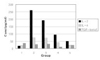
Bacterial extracts were evaluated for their ability to suppress the production of cytokine of lymphocyte to mitogens of PHA. Data of Table 1 and Figure 1 are the representative results of experiments. Results are the mean values of six samples of duplicate cultures. PHA-activated group (Group 2) exhibited higher level of IL-2 and IL-4 than medium only control group (Group 1) (p < 0.05). As compared with PHA-activated (Group 2), high and medium concentration of SEF (Group 4, 5) decreased the production of IL-2 and IL-4 from lymphocyte (p < 0.05), but there was no statistically significant difference between low concentration of SEF (Group 3) and PHA-activated group (Group 2). So we can suggest that SEF cause a dose-dependent reduction in PHA-induced cytokine production of IL-2, and IL-4. But the production of TGF-β1 was independent of concentration of SEF (p > 0.05).
IV. Discussion
Microbial products represent an important source of immunoregulatory agents. In particular, several microorganisms are capable of suppressing the immune response through various products, including toxins, enzymes, cell wall components, and metabolites. These immunosuppressive products may alter the immune system via different mechanisms. In some instances these agents indirectly modify lymphocyte response by directly affecting monocytes or macrophage activities directly. For example, Spirochetes inhibited lymphocytes function and Leishmania donovani impaired the synthesis and release of IL-1 from macrophages17,18). Sonicated extract from Prevotella intermedia, Porphylomonas asaccharolytica, Porphyromonas endodontalis and Prevotella melaninogenica were capable of suppressing human T- and B-cell response in dose-dependent manner. The immunosuppressive activity is nondialyzable and heat labile19).
This study showed that the level of IL-2 and IL-4 in lymphocytes stimulated with PHA were significantly higher than those of the negative control group. So, PHA can have sufficient mitogenic effect to lymphocytes. But sonicated extracts of E. faecalis suppressed in vitro IL-2, IL-4 production of human lymphocyte in a dose-dependent manner. Ability of E. faecalis to impair the production of IL-2 and IL-4 may be related to the suppression of T-cell activation. In our previous studies, in contrast, lipopolysaccharide of Porphyromonas endodontalis in vitro stimulated the production of macrophage inflammatory protein (MIP)-1 alpha and MIP-1 beta from human polymorphonuclear leukocyte20). In rat pulpitis experimentally induced by specific bacteria, Streptococcus mutans, Porphylomonas endodontalis stimulated the production of Interferon-γ, IL-2 and IL-414). These studies showed that P. endodontalis have inflammatory effects by stimulation, but in other reports, sonic extract of P. endodontalis in lymphocyte in vitro have immunosuppressive effects by inhibition19), and in this study E. faecalis inhibited the cytokine production of lymphocytes. Much further investigation is required to clarify the stimulation and inhibition of cytokines. In contrast, there was no significant difference in the level of TGF-β1 in regardless of SEF, this results show that TGF-β1 is released by other mechanism.
When comparing with the level of each cytokine, Results showed that the level of IL-2 predominated over those of other cytokine IL-4, and TGF-β1. Considering the concentration of IL-2, Th1 lymphocytes may be dominant in human peripheral blood lymphocytes.
It has been proposed that impaired host defense may play a pivotal role in the pathogenesis of many infections. The data presented in this study suggest that microbial mediated immunosuppression may contribute to the pathogenesis of endodontic infection by altering the nature and consequences of host-parasite interactions21). Microbial virulence may be the consequence of several properties, including the ability of certain species to resist, escape, or pervert host defense mechanism. Some causative virus infects and destroys a subpopulation of T lymphocytes, such as human immunodeficiency virus. Such inhibitory factors could lead to a state of immunological hyporesponsiveness that favors colonization by the initiating organism or by other opportunistic organisms. Our current finding that E. faecalis inhibits the immune response suggests that impaired host defense mechanism may contribute to the disease process.
This results suggested that bacterial protein sonicated extract of E. faecalis can inhibit the immune response by lymphocyte and this could be one of possible factors why E. faecalis are found frequently in the teeth with failed endodontic treatment.
V. Conclusion
According to this study we could summarize as follows: PHA activated expression of IL-2 and IL-4 from lymphocytes compared with negative control group (p < 0.05). As compared with PHA-activated group, high and medium concentration of SEF decreased the production of IL-2 and IL-4 from lymphocytes (p < 0.05). There was no statistically significant difference between low concentration of SEF and PHA-activated group. The production of TGF-β1 was independent of concentration of SEF (p > 0.05). So we can suggest that SEF cause a dose-dependent reduction in PHA-induced cytokine production of IL-2, and IL-4.

 XML Download