

 PDF
PDF ePub
ePub Citation
 Print
Print


Abstract
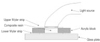
The objectives of this study was to evaluate current visible light curing units regarding microhardness and microleakage. Fourty samples of composite resin(Z-250, 3M) were cured by different light curing units(Flipo, LOKKI; Credi II, 3M; XL 3000, 3M; Optilux 500,Demetron) in acrylic blocks. Microhardness was measured using a calibrated Vickers indenter on both top and bottom surfaces after 24 hours of storage in air at room temperature. Class V cavities were prepared on buccal and lingual surfaces of fourty extracted human molars. Each margin was on enamel and dentin/cementum. Composite resin(Z-250, 3M) was filled in cavities and cured by four different light curing units(Flipo, LOKKI; Credi II, 3M; XL 3000, 3M; Optilux 500, Demetron).
The results of this syudy were as follows:
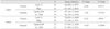
Microhardness
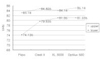
1. Flipo showed low microhardness compared to Optilux 500, Credi II significantly in upper surface. Flipo didn't show a significant difference compared to XL 3000.
2. The microhardness resulting from curing with Flipo was lower than that of others on lower surfaces.
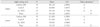
Microleakage
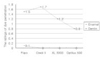
1. Dentin margin showed significantly high dye penetration rate than enamel margin in all groups(p<0.05).
2. No significant differences were found on both enamel and dentin margin regarding curing units.


References
1. Sakaguchi RL, Peters MC, Nelson SR, Douglas WH, Poort HW. Effects of polymerization contraction in composite restorations. J Dent. 1992. 20:178–182.

2. Gerzina TM, Hume WR. Effect of hydrostatic pressure on the diffusion of monomers through dentin in vitro. J Dent Res. 1995. 74:369–373.
3. Geurtsen W. Substances released from dental resin composites and glass ionomer cements. Eur J Oral Sci. 1998. 106(2 Pt 2):687–695. Review.
4. Hamid A, Okamoto A, Iwaku M, Hume WR. Component release from light-activated glass ionomer and compomer cements. J Oral Rehabil. 1998. 25:94–98.
5. Vargas MA, Cobb DS, Schmit JL. Polymerization of composite resins: argon laser vs conventional light. Oper Dent. 1998. 23:87–93.
6. Venhoven BA, de Gee AJ, Davidson CL. Polymerization contraction and conversion of light-curing bis GMA-based methacrylate resins. Biomaterials. 1993. 14:871–875.
7. Davidson CL, de Gee AJ. Relaxation of polymerization contraction stresses by flow in dental composites. J Dent Res. 1984. 63:146–148.
8. Tarle Z, Meniga A, Ristic M, Sutalo J, Pichler G, Davidson CL. The effect of the photopolymerization method on the quality of composite resin samples. J Oral Rehabil. 1998. 25:436–442.
9. Mehl A, Hickel R, Kunzelmann KH. Physical properties and gap formation of light-cured composites with and without 'softstart-polymerization'. J Dent. 1997. 25:321–330.
10. Uno S, Asmussen A. Marginal adaptation of a restorative resin polymerized at reduced rate. Scand J Dent Res. 1991. 99:440–444.
11. Tirtha R, Fan PL, Dennison JB, Powers JM. In vitro depth of cure of photo-activated composite. J Dent Res. 1982. 61:1184–1187.
12. Sturdevant CM. The art and science of operative dentistry. 1995. 3rd ed. St Louis: Mosby;260.
13. Rueggeberg FA, Caughman WF, Curtis JW Jr. Effect of light intensity and exposure duration on cure of resin composite. Oper Dent. 1994. 19:26–32.
14. Curtis JW, Rueggeberg FA, Lee AJ. Curing efficiency of the turbo tip. Gen Dent. 1995. 43:428–433.
15. Loney RW, Price RBT. Temperature transmission of high-output light-curing units through dentin. Oper Dent. 2001. 26:516–520.
16. Bouschlicher MR, Heiner AD. Polymerization shrinkage force with xenon short arc or QTH photoillumination. J Dent Res. 2001. 80(Special Issue):253. Abstract #1737.
17. Haitz RH, Craford MG, Wiessman RH. Handbook of optics. 1995. vol 2. New York: McGraw Hill;12.1–12.9.
18. Nakamura S, Mukai T, Senoh M. Candela-class high brightness InGaN/AlGaN double heterostructure blue-light-emitting diodes. Appl Phys Lett. 1994. 64:1687–1689.
19. Rueggeberg FA, Craig RG. Correlation of parameters used to estimate monomer conversion in a light-cured composite. J Dent Res. 1988. 67:932–937.
20. Rueggeberg FA, Caughman WF, Curtis JW Jr. Factors affecting cure at depths within light-activated resin composite. Am J Dent. 1993. 6:91–95.
21. Yearn JA. Factors affecting cure of visible light activated composites. Int Dent J. 1985. 35:218–225.
22. McCabe JF, Carrick TE. Output from visible-light activation units and depth of cure of light-activated composite. J Dent Res. 1989. 68:1534–1539.
23. Price RB, Dérand T, Loney RW, Andreou P. Effect of light source and specimen thickness on the surface hardness of resin composite. Am J Dent. 2002. 15:47–53.
24. Cook WD. Spectral distribution of dental photopolymerizing sources. J Dent Res. 1982. 61:1436–1438.
25. Park SH, Krejci I, Lutz F. Microhardness of resin composites polymerized by plasma arc or conventional visible light curing. Oper Dent. 2002. 27:30–37.
26. Gagliani M, Fadini L, Ritzmann JM. Depth of cure efficacy of high-power curing devices vs traditional halogen lamps. J Adhes Dent. 2002. 4:41–47.
27. Sharkey S, Roy N, Burke F, Ziada H, Hannigun A. Surface hardness of light-activated resin composites cured by two different visible-light sources:An invitro study. Quintessence Int. 2001. 32:401–405.
28. Johnston W, Leung R, Fan P. Amathematical model for post-irradiation hardening of photoactivated composite resins. Dent Mater. 1985. 1:191–194.
29. Martin FE. A survey of the efficiency of visible light curing units. J Dent. 1998. 26:239–243.
30. Mitton BA, Wilson NHF. The use and maintenance of visible light activating units in general practice. Br Dent J. 2001. 191:82–86.
31. Sakaguchi RL, Douglas WH, Peters MC. Curing light performance and polymerization of composite restorative materials. J Dent. 1992. 20:183–188.
32. Solomon CS, Osman YI. Evaluating the efficacy of curing lights. SADJ. 1999. 54:357–362.
33. Miyazaki M, Hattori T, Ichiishi Y, Kondo M, Onose H, Moore B. Evaluation of curing units used in dental office. Oper Dent. 1998. 23:50–54.
34. Leonard DL, Charlton DG, Roberts HR, Hilton TJ, Zionic A. Determination of the minimum irradiance required for adequate polymerization of a hybrid and a microfill composite. Oper Dent. 2001. 26:176–180.
35. Prati C, Chersoni S, Montebugnoli L, Montanari G. Effect of air, dentin and resin-based composite thickness on light intensity reduction. Am J Dent. 1999. 12:231–234.
36. Leonard DL, Charlton DG, Hilton TJ. Effect of curing-tip diameter on the accuracy of dental radiometers. Oper Dent. 1999. 24:31–37.
37. Rueggeberg FA, Jordan DM. Effect of light-tip distance on polymerization of resin composite. Int J Prosthodont. 1993. 6:364–370.
38. Rueggeberg FA, Caughman WF, Curtis JW Jr, Davis HC. Factors affecting cure at depths within light-activated resin composites. Am J Dent. 1993. 6:91–95.
39. Venhoven BAM, De Gee AJ, Davidson CL. Polymerization contraction and converson of light-curing Bis-GMA-based methacrylate Resins. Biomaterials. 1993. 14:871–875.
40. Brackett WW, Haisch LDA, Covey DA. Effect of plasma arc curing on the microleakage of Class V resin-based composite restorations. Am J Dent. 2000. 13:121–123.
41. Feilzer AJ, Dooren LH, de Gee AJ, Davidson CL. Influence of light intensity on polymerization shrinkage and integrity of restoration-cavity interface. Eur J Oral Sci. 1995. 103:322–326.
42. Sakaguchi RL, Peters MC, Nelson SR, Douglas WH. Effects of polymerization contraction in composite restorations. J Dent. 1992. 20:178–182.
43. Peutzfeldt A, Sahafi A, Asmussen E. Characterization of resin composites polymerized with plasma arc curing units. Dent Mater. 2000. 16:330–336.
44. Uno S, Asmussen E. Marginal adaptation of a restorative resin polymerized at reduced rate. Scand J Dent Res. 1991. 99:440–444.
45. Rueggeberg F. Contemporary issues in photocuring. Compend Contin Educ Dent Suppl. 1999. (25):S4–S15.
46. Hofmann N, Hugo B, Schubert K, Klaiber B. Comparison between a plasma arc light source and conventional halogen curing units regarding flexural strength, modulus and hardness of photoactivated resin composites. Clin Oral Investig. 2000. 4:140–147.
47. Davidson-Kaban SS, Davidson CL, Feilzer AJ. The effect of curing light variations on bulk curing and wall-to-wall quality of two types and various shades of resin composite. Dent Mater. 1997. 13:344–352.
48. Ferracane JL. Correlation between hardness and degree of conversion during the setting reaction of unfilled dental restorative resins. Dent Mater. 1985. 1:11–14.
49. Leonard DL, Charlton DG, Hilton TJ. Effect of curing-tip diameter on the accuracy of dental radiometers. Oper Dent. 1999. 24:31–37.
 XML Download