Journal List > J Korean Acad Conserv Dent > v.29(6) > 1056137

Kim, Lee, Cho, Son, Kim, Seok, and Um: Dentin bond strength of bonding agents cured with Light Emitting Diode

ABSTRACT

This study compared the dentin shear bond strengths of currently used dentin bonding agents that were irradiated with an LED (Elipar FreeLight, 3M-ESPE) and a halogen light (VIP, BISCO). The optical characteristics of two light curing units were evaluated. Extracted human third molars were prepared to expose the occlusal dentin and the bonding procedures were performed under the irradiation with each light curing unit. The dentin bonding agents used in this study were Scotchbond Multipurpose (3M ESPE), Single Bond (3M ESPE), One-Step (Bisco), Clearfil SE bond (Kuraray), and Adper Prompt (3M ESPE). The shear test was performed by employing the design of a chisel-on-iris supported with a Teflon wall. The fractured dentin surface was observed with SEM to determine the failure mode.
The spectral appearance of the LED light curing unit was different from that of the halogen light curing unit in terms of maximum peak and distribution. The LED LCU (maximum peak in 465 ㎚) shows a narrower spectral distribution than the halogen LCU (maximum peak in 487 ㎚). With the exception of the Clearfil SE bond (P < 0.05), each 4 dentin bonding agents showed no significant difference between the halogen light-cured group and the LED light-cured group in the mean shear bond strength (P > 0.05).
The results can be explained by the strong correlation between the absorption spectrum of cam-phoroquinone and the narrow emission spectrum of LED.

References

1. Council on Dental Materials, Instruments, and Equipment. Visible light-cured and activating units. J Am Dent Assoc. 110(1):100–102. 1985.
2. Blankenau RJ, Kelsey WP, Powell GL, Shearer GO, Barkmeier WW, Cavel WT. Degree of composite resin polymerization with visible light and argon laser. Am J Dent. 4(1):40–42. 1991.
3. Ferracane JL, Mitchem JC, Condon JR, Todd R. Wear and marginal breakdown of composites with various degrees of cure. J Dent Res. 76(8):1508–1516. 1997.
crossref
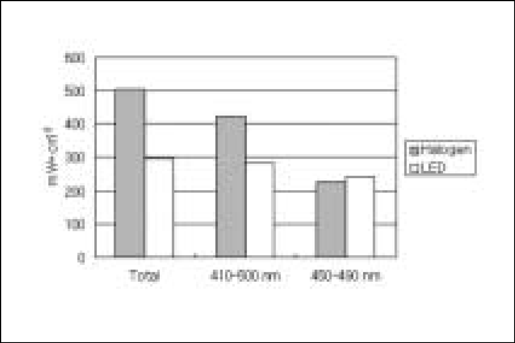
4. Rueggeberg FA, Twiggs SW, Caughman WF, Khajotia S. Life-time intensity profiles of 11 light-curing units. J Dent Res. 75:380 Abstr.. (No. 2897):1996.
5. Friedman J. Variability of lamp characteristics in dental curing lights. J Esthet Dent. 1(6):189–190. 1989.
crossref
6. Martin FE. A survey of the efficiency of visible light curing units. J Dent. 26(3):239–243. 1998.
crossref
7. Barghi N, Berry T, Hatton C. Evaluating intensity output of curing lights in private dental offices. J Am Dent Assoc. 125(7):992–996. 1994.
crossref
8. Miyazaki M, Hattori T, Ichiishi Y, Kondo M, Onose H, Moore BK. Evaluation of curing units used in private dental offices. Oper Dent. 23(2):50–54. 1998.
9. Mills RW. Blue light emitting diodes: Another method of light curing? Br Dent J. 178:169. 1995.
10. Nakamura S, Mukai T, Senoh M. Candela-class high brightness InGaN/AlGaN double heterostructure bluelight-emitting diodes. Appl Phys Lett. 64:1687–1689. 1994.
11. Duke ES. Light-emitting diodes in composite resin photopolymerization. Compend Contin Educ Dent. 22(9):722–725. 2001.
12. Yap AU, Soh MS. Thermal emission by different light-curing units. Oper Dent. 28(3):260–266. 2003.
13. Fujibayashi K, Ishimaru K, Takahashi N, Kohno A. Newly developed curing unit using blue light-emitting diodes. Dent Jap. 34:49–53. 1998.
14. Mills RW, Jandt KD, Ashworth SH. Dental composite depth of cure with halogen and blue light emitting diode (LED) technology. Br Dent J. 186(8):388–391. 1999.
15. Stahl F, Ashworth SH, Jandt KD, Mills RW. Light-emitting diode (LED) polymerization of dental composites: flexural properties and polymerization potential. Biomaterials. 21(13):1379–1385. 2000.
16. Mills RW, Uhl A, Jandt KD. Optical power outputs, spectral and dental composite depths of cure, obtained with blue light emitting diode (LED) and halogen light curing units (LCUs). Br Dent J. 193(8):459–463. 2002.
17. Hansen EK, Asumussen E. Reliability of three dental radiometers. Scand J Dent Res. 101(2):115–119. 1993.
18. Leonard DL, Charlton DG, Hilton TJ. Effect of curing-tip diameter on the accuracy of dental radiometers. Oper Dent. 24(1):31–37. 1999.
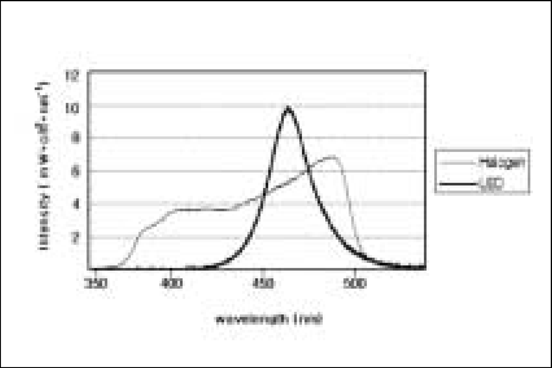
19. Cook WD. Spectral distribution of dental photopolymerization sources. J Dent Res. 61:1436–1438. 1982.
20. McCabe JF, Carnick TE. Output from visible-light activation units and depth of cure of light-activated composites. J Dent Res. 68(11):1534–1539. 1989.
crossref
21. Nomoto R. Effect of light wavelength on polymerization of light-cured resins. Dent Mater J. 16(1):60–73. 1997.
crossref
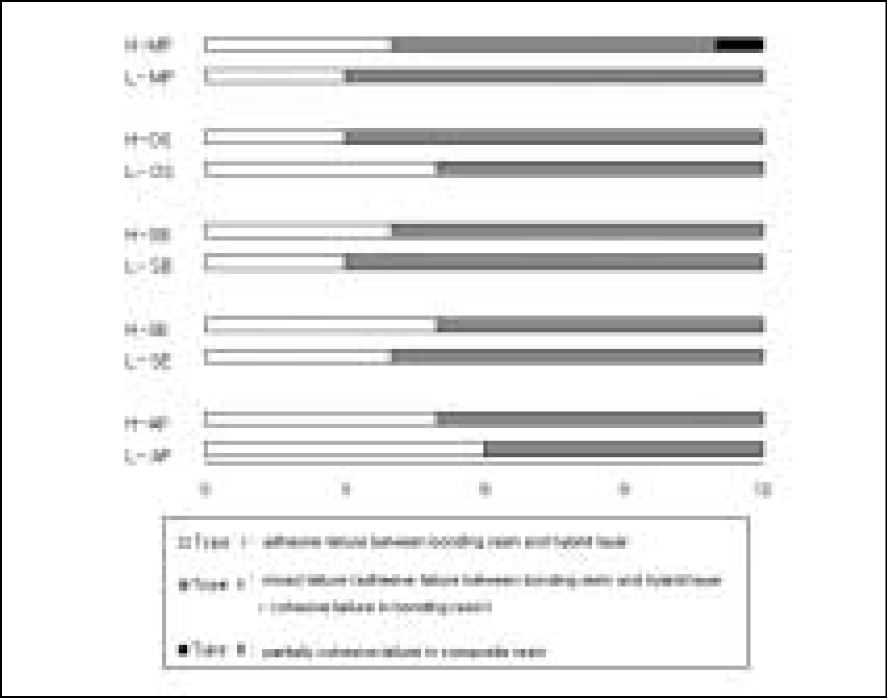
22. Dickens SH, Milos MF. Relationship of dentin shear bond strengths to different laboratory test designs. Am J Dent. 15(3):185–192. 2002.
23. Versluis A, Tantbirojn D, Douglas WH. Why do shear bond tests pull out dentin? J Dent Res. 76(6):1298–1307. 1997.
crossref
24. Stansbury JW. Curing dental resins and composites by photopolymerization. J Esthet Dent. 12:300–308. 2000.
crossref
25. Hayakawa T, Kikutake K, Nemoto K. Effectiveness of the addition of water-soluble photoinitiator into the self-etching primers on the adhesion of a resin composite to polished dentin and enamel. Dent Mater J. 18(3):324–333. 1999.
crossref
26. Burgess JO, DeGoes M, Walker R, Ripps AH. An evaluation of four light-curing units comparing soft and hard curing. Pract Periodontics Aesthet Dent. 11(1):125–132. 1999.
27. Koliniotou-Kubia E, Jacobsen PH. The effect of irradi-antion time on the physicalperoperties of light-cured resins. Clin Mater. 6(1):21–28. 1990.
28. Takahashi A, Sato Y, Uno S, Pereira PN, Sano H. Effects of mechanical properties of adhesive resins on bond strength to dentin. Dent Mater. 18(3):263–268. 2002.
crossref

Figure 1.
Schematic diagram of the shear bond strength test used in this study.
  1. Embedding resin

  2. Teflon-coated metal iris

  3. Teflon wall

  4. Composite

  5. Load application chisel

jkacd-29-504f1.tif
Figure 2.
Spectral distributions of the two LCUs.
jkacd-29-504f2.tif
Figure 3.
Spectral intensity of two LCUs used in this study.
jkacd-29-504f3.tif
Figure 4.
The failure mode.
jkacd-29-504f4.tif
Figure 5.
Adhesive failure.
jkacd-29-504f5.tif

a. Total fractured surface on the dentin side of a H-SE specimen (× 23). b. Higher magnification of the sections of the fractured surface marked (b) in figure 6a (× 400). c. Higher magnification of the sections of the fractured surface marked (c) in figure 6b (× 2000). d. Higher magnification of the sections of the fractured surface marked (d) in figure 6c (× 20,000). Shear force is suggested by the appearance of the fractured resin tag.

Figure 6.
Mixed failure; adhesive failure + cohesive failure within the DBA.
jkacd-29-504f6.tif

a. Total fractured surface on the dentin side of a L-SB specimen (× 23). b. Higher magnification of the fractured surface marked (b) in figure 7a (× 400). c, d Higher magnification of the sections of the fractured surface marked (c), (d) in figure 7b (× 1000). Figure 7c shows cohesive failure of the bonding resin. Figure 7d shows adhesive failure between the hybrid layer and the bonding resin.

Figure 7.
Partially cohesive failure within the composite.
jkacd-29-504f7.tif

a. Total fractured surface on the dentin side of a H-MP specimen (× 23). Partially cohesive failure in the composite was observed in the upper portion. b. Higher magnification of the sections of the fractured surface marked (b) in figure 8a (× 400). c, d. Higher magnification of the sections of the fractured surface marked (c), (d) in figure 8b (× 3000).

Table 1.
Manufacturers, components and bonding procedures of the different dentin bonding systems and composites used in this study.
Bonding systems Components Bonding procedures Composite resin
Scotchbond Multipurpose 3M ESPE (MP) Primer : HEMA, polyalkenoic acid copolymer, water Adhesive : Bis-GMA, HEMA, initiator, amine Etched with 35% phosphoric acid for 15s, rinsed, lightly dried with a gentle air stream to leave a moist surface, primer applied, dried with gentle air stream, adhesive applied, light-cured for 10s. Z-100 (A2 shade)

Single Bond 3M ESPE (SB) HEMA, Bis-GMA, dimethacrylates, polyalkenoic acid copolymer, photoinitiator, ethanol, water Etched with 35% phosphoric acid for 15s, rinsed, lightly dried with a gentle air stream to leave a moist surface, adhesive applied in two consecutive coats, air-dried, light-cured for 10s Z-100 (A2 shade)

One-Step Bisco (OS) HEMA, BPDM, Bis-GMA, photoinitiator, acetone Etched with 32% phosphoric acid for 15s, rinsed, lightly dried to leave the dentin moist, two coats of adhesive applied with agitation, air dried, light-cured for 10 s Renew (A2 shade)

Clearfil SE Bond Kuraray (SE) Primer: MDP,HEMA, hydrophilic DMA, CQ, N,N-Diethanol-p-toluidine, water Adhesive : MDP, Bis-GMA, hydrophobic dimethacrylate, HEMA, CQ, toluidine, silanated colloidal silica Dried with light air, SE primer applied, wait 20s, evaporate with a light air flow, adhesive applied, gently air blow, light-cured for 10s Clearfil AP-X (A2 shade)

Adper Prompt 3M ESPE (AP) Liquid A: methacrylated phosphoric ester, Bis-GMA, initiators based on CQ, stabilizer Liquid B : water, HEMA, polyalkenoic acid, stabilizer Aggressively mix each one drop from solution A and solution B for 5 s. the mixed adhesive applied and rubbed in the solution with a moderate finger pressure for 15s. gently dried, light-cured for 10s Z-100 (A2 shade)

Abbreviation: Bis-GMA, bisphenyl-glycidyl-methacrylate; HEMA, 2-hydroxyethyl methacrylate; MDP, 10-methacryloy-loxydecyl dihydrogen phosphate; BPDM, Biphenyl Dimethacrylate; DMA, Dimethacrylate; CQ, Camphoroquinone

Table 2.
The groups examined in this study; Abbreviation.
Halogen (VIP) LED (Elipar Freelight)
Scotchbond Multi-Purpose H-MP L-MP
Single Bond H-SB L-SB
ONE-STEP H-OS L-OS
Clearfil SE Bond H-SE L-SE
Adper Prompt H-AP L-AP
Table 3.
Light power measurement and conversion to intensity.
Manufacturer Demetron EPM 1000 Intensity calculated
(㎽ ㎝-2) (㎽ ㎝-2) (㎽) (㎽ ㎝-2)
VIP (Halogen) 400 350 254 505
Elipar Freelight (LED) About 400 310 148 294
Table 4.
Mean shear bond strength values of the bonding agents (MPa) irradiated with the halogen (VIP) and LED (Elipar Freelight).
DBA Halogen LED p-value
mean ± s.d mean ± s.d
SE 37.60 ± 7.34 a 25.38 ± 9.24 A, B 0.002 *
SB 25.32 ± 8.63 b 26.98 ± 5.37 A 0.577
MP 24.24 ± 8.06 b 22.22 ± 7.62 A, B 0.535
OS 20.91 ± 8.02 b 21.29 ± 5.62 A, B 0.892
AP 20.14 ± 3.69 b 16.81 ± 5.21 B 0.085

The same letters indicate no significant difference between the groups.

* Means a statistically significant difference.

TOOLS
Similar articles