

 PDF
PDF ePub
ePub Citation
 Print
Print


I. INTRODUCTION
Treponema denticola has long been considered as a periodontal pathogen1). Listgarten also reported that spirochetes are conspicuous inhabitants of subgingival plaque in patients with gingivitis and periodontitis2). Although this bacteria is well known to be important periodontal pathogens, several studies suggest that spirochetes may be involved in pulpal and periapical infections as well. For instance, Thilo et al.3) reported that the root canal flora of decayed teeth contained as much as 6% of spirochetes. Likewise, Nair4) reported that the spirochetes form a significant component of the flora of periapical specimen. Recent studies5,6) using 16S rDNA-directed polymerase chain reaction (PCR) technique confirmed that the occurrence of Treponema denticola accounted for 52.4% of the flora in infected root canals and 50% in acute alveolar abscesses of endodontic origin. Thus, spirochetes or their byproducts may contribute to the inflammatory process and pathogenesis of the periradicular infection. However, it is not clear how spirochetes are involved in the etiology of the disease. In this regard, Shenker et al.7) demonstrated that soluble sonic extracts of several strains of T. denticola inhibit human peripheral blood lymphocyte (HPBL) proliferative responses to both mitogen and antigens in vitro with no effect on cell viability. They have also observed that these effects are due to a protein compound of two polypeptides of 50 and 56 kDa.
Among the lymphocyte populations in the host defense system, T cell has been well known to play an important role in periapical lesion development and maintenance. 60% of peripheral T cells are CD4 positive T helper cells and these cells are subdivided into Th1 and Th2 cells. Due to the lack of definitive phenotypic markers for these subsets, cytokine production can be a good indicator for their activities in response to bacterial stimulations8). In fact, Interleukin-2 (IL-2) and Interleukin-4 (IL-4) are the major effector cytokines released from Th1 and Th2, respectively. IL-2 stimulates T cells proliferation and involved in cell mediated immunity while IL-4 promotes the B cell to differentiate into plasma cell that produces antibodies and thus mediating humoral immunity9).
Recent studies10-13) have investigated that the suppression of lymphocyte proliferation by pathogenic bacteria related to endodontic treatment failures. Furthermore, Lee et al.14) clearly showed that immunoinhibitory protein extracted from T. denticola suppressed proliferative ability of lymphocytes by arresting cell cycle. However, studies on the effects of T. denticola on cytokine-producing function of lymphocytes are lacking. Therefore, the objective of this study was to compare the IL-2 and IL-4 expression by human T cells before and after the addition of sonicated T. denticola extract.
II. MATERIALS AND METHODS
1. T lymphocyte preparation
T lymphocytes were prepared from 20ml of EDTA-anticoagulated venous blood of healthy donors. Cells were isolated by buoyant density centrifugation on Ficoll-Hypaque (Pharmacia LKB Biot-echnology, Piscataway, NJ, USA). T Cells were then washed twice with Hanks' balanced salt solution, centrifuged at 16,000 × g for 10 min at 4℃, and diluted to 2 × 10⁶ viable cells per ml culture medium consisting of RPMI 1640, 2% penicillin/streptomycin, and 2% fetal bovine serum.
2. Sonicated bacterial extracts preparation
Treponema denticola strain LL2513 was grown in 500ml of TYGVS (Trypticase, yeast extract, glucose, volatile fatty acid, serum) spirochete medium containing veal infusion broth (Difco Laboratories, Detroit, MI, USA) supplemented with yeast extract (BBL), trypticase peptone, ammonium sulfate, L-cycteine hydrochloride, glucose, and volatile fatty acid solution (acetic acid, propionic acid, n-butyric acid, isovaleric acid and D,L-methylbutyric acid in dH2O). Anaerobic cultures of T. denticola were grown at 37℃ in 20-ml glass tubes. After 72 h, bacterial cell suspensions were harvested by centrifugation at 16,000 × g for 15 minutes at 4℃ and washed 3 times in Phosphate buffered saline (PBS) containing PMSF. The washed bacterial cells were lyophilized and sonicated on ice with a Sonic Dismembrator (Model 550, Fisher Scientific, Pittsburg, PA, USA) for a total of 7 min with 30-s pulses. Disruption of the bacterial cell was confirmed microscopically and these sonicates were collected and centrifuged at 85,000 × g for 60 minutes. The supernatant was dialyzed against PBS, and the protein that remained in suspension was designated as sonicated T. denticola extracts.
3. Sonicated extracts response experiment
For the experiment, T cell suspension containing 1 × 10⁶ cells was placed into each well of flat-bottomed 24-well plate. Each culture received medium (RPMI supplemented with 100U of penicillin/ml, 100mg of streptomycin and 2% fetal bovine serum) or optimal concentrations (12.5 µg/ml) of sonicated extracts diluted in medium. Additionally, all experimental cultures received 100 µl of phytohemagglutinin (PHA; Sigma Chemical Co., St. Louis, MO, USA) as an antigenic stimulant. Experimental groups were arranged in 3 groups: Group 1 was designated untreated control cells; group 2 consisted of cells with PHA activation alone;; group 3 contained cells pretreated with 12.5 µg/ml of sonicated extracts and PHA activation.
4. Cytokine assay
After 72h incubation, the amount of IL-2 and IL-4 present in the culture supernatants were assessed by Enzyme-linked immunosorbent assay (Quantikine colorimetric sandwich ELISA kits; R&D System, Inc., Minneapolis, MN, USA) according to the manufacturer's protocol. Briefly, monoclonal antibodies specific for IL-2 and IL-4 were precoated onto enzyme-linked immunosorbent assay plate. Dilutions of standards and culture supernatants were then added to individual wells in duplicate. After any unbound substances were washed away, an enzyme-linked polyclonal antibody specific for IL-2 and IL-4 was added to the wells. After a further wash to remove any unbound antibody-enzyme reagent, a substrate solution was added to the wells, and the color was developed. The optical density was read at an absorbance of 450 nm, and the cytokine concentration was quantified from the standard curve. Three samples in each group were analyzed in duplicate.
III. RESULTS
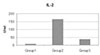
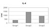
The means and standard deviations of IL-2 and IL-4 expressions are summarized in Table 1.
Compared to the control (Group 1), T lymphocytes were well responded to the PHA stimulation. As shown in Table 1, PHA activated cells (Group 2) exhibited high levels of both IL-2 and IL-4. These cytokine productions were significantly suppressed when the cells were pre-exposed to 12.5 µg/ml (Group 3) of sonicated T. denticola extracts (p < 0.05) (Figure 1, 2).
IV. DISCUSSION
Treponema denticola have been rarely found associated with endodontic diseases, however, unidentified spirochete species have now been found in endodontic infections by employing polymerase chain reaction (PCR) method6,15-17). In this regard, Smallwood et al.18) recently reported the occurrence of spirochetes in all samples collected from infected canals using PCR. Likewise, Siqueira et al.5) demonstrated that Treponema denticola was detected 11 of 21 infected root canal cases.
T. denticola is a gram-negative, anaerobic, helically shaped, highly motile bacteria. It has an assembly of virulence factors that can contribute to its pathogenicity. T. denticola can adhere to diverse host cells and other tissue components and it has also been demonstrated to invade cells and tissues. In addition, it may have immunomodulatory effect, including a number of specific effects on polymorphonuclear leukocytes and lymphocytes. Some T. denticola surface-expressed proteins have adhesion, cytotoxic and tissue destructive activities, including the major surface protein and the chymotrypsin-like protease complex. Other extracellular membrane-associated proteolytic and hydrolytic enzymes, short chain fatty acids and volatile sulfur compounds are other potentially virulence factors of oral spirochetes19-20).
There are accumulating evidences that pathogenic bacteria associated with endodontic infection and have been shown to alter the host defense system. For example, Yoshida et al.13) reported that sonicated material from Fusobacterium nucleatum can either suppress or stimulate T cell proliferation in the presence of dental pulp accessory cells. Likewise, Gentry-Weeks et al.21) reported that Enterococcus faecalis can survive in mouse peritoneal macrophages. Recent study of Lee et al.12) demonstrated that sonicated Enterococcus faecalis extracts inhibit lymphocyte proliferation by arresting cell cycle progression. Thus, this immunosuppressive process is relevant to the pathogenesis of the periapical disease. This kind of immunosuppression by bacterial byproducts may act differently by interfering with either the induction or the expression of immune reaction. Sometimes these immunomodulatory agents can activate T cells, sometimes can suppress T cells, or have direct effect on both precursor and mature effector cells.
In this regard, Shenker et al.7) have previously shown that extracts of the oral spirochete, T. denticola, contain an immunosuppressive protein which impairs human lymphocyte proliferation. Recently, Lee et al.14) confirmed this immunoinhibitory effect by inducing irreversible G1 arrest in activated human lymphocytes. Spirochetes may contribute to the pathogenesis of a member of disorders including periodontal and periradicular diseases; however, the mechanism by which these organisms act to cause the disease is unknown.
In the present study, we have investigated whether the sonicated T. denticola extracts have the immunosuppressive effect on cytokine-producing function of T cells. As shown in data, PHA stimulation has sufficient antigenic effect on the T cells (Group 2 in Table 1). The levels of both IL-2 and IL-4 expressions were significantly increased when cells were activated with PHA (Group 2), compared to those of the unstimulated control (Group 1). These cytokine productions were down-regulated when T cells were exposed to the sonicated T. denticola extracts prior to PHA activation (Group 3). This suppression of cytokine expression indicated that the immunoinhibitory protein extracted from T. denticola has the ability of inhibiting lymphocyte function. This impaired lymphocyte function, in turn, would adversely affect the development of normal immunologic defense mechanism and as a result, it contributes the pathogenesis of periapical lesion.
T helper cells actively participate in immune defense via producing cytokines in response to the antigenic stimulation. Mediators produced by subset Th1 cells increase inflammation and bone resorption while Th2 cell-derived cytokines are inhibitory22). Th1 cells initiate cell-mediated immune reactions whereas Th2 cells orchestrate humoral immune responses. The balance between Th1 and Th2 cell is critical in periapical lesion development and thus, protects hard tissue from direct bacterial invasion9). Therefore, Bacteria-induced reduction or hypo-production of selected cytokines derived from the Th1 and Th2 cell is responsible for the host tissue destruction.
In conclusion, the cytokine-producing functions of both Th1 and Th2 cells were significantly perturbed by sonicated T. denticola extracts. This observation suggests that the T. denticola induced-immunosuppression in T cell function could be a part of the pathogenic mechanism of the periapical lesion.

 XML Download