

 PDF
PDF ePub
ePub Citation
 Print
Print


Abstract
The purpose of this study is to compare and to evaluate the effects of the degree of saturation on the progression of artificial root caries lesion.
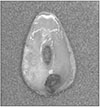
A total of 8 human premolars without any defects and cracks selected and the cementum were removed and the teeth were cleaned with ultrasonic device and pumice without fluoride.
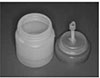
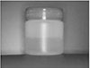
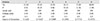
Each tooth was sectioned into 6 pieces and they were ground with #800 sandpaper until they had a thickness of 200µm. Specimens were applied with nail vanish except for the 2-3 mm window area after application of bonding agent. Under the constant pH, the specimens were divided into 6 groups (degree of saturation; 0.1415, 0.1503, 0.1597, 0.1676, 0.1771, 0.1977). Each group was immersed in acid buffer solution for 1, 2, 3, 5 days under controlled temperature (25℃) and imbibed in water and examined using the polarizing microscope.
The results were as follows
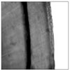
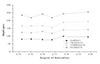
1. Although the degree of saturation of demineralization solution decreased, the depth of penetration in the dentin was constant.
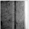
2. Erosion was observed on the surface of all the teeth in the group I, II. In the group III, IV, V, surfaces were not changed. The teeth in the group VI showed the more mineralized surface but not the shape of the dentinal tubules distinctively.
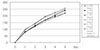
3. In all groups, the lesion progressed rapidly at the first day of the experiment, but increased gradually as time elapsed.
Figures and Tables


References
1. Christensen GJ. A new challenge-root caries in mature people. J Am Dent Assoc. 1996. 127:379–380.

2. Shay K. Root caries in the older patient. Dent Clin North Am. 1997. 41:763–793.
3. Katz RV, Hazen SP, Chilton NW, Mumma RD. Prevalence and intraoral distribution of root caries in an adult population. Caries Res. 1982. 16:265–271.
4. Joshi A, Papas AS, Giunta J. Root caries incidence and associated risk factors in middle-aged and older adults. Gerodontology. 1993. 10:83–89.
5. Hazen SP, Chilton NW, Mumma RD JR. The problem of root caries I; Literature review and clinical description. JADA. 1973. 86:137–144.
6. Banting DW, Ellen RP, Fillery ED. Prevalence of root caries among institutionalized older persons. Community Dent Oral Epidemiol. 1980. 8:84–88.
7. Sturdevant CM, Roberson TM, Heymann HO, Sturdevant JR. The art and science of oerative dentistry. 1995. 3rd Ed. St Louis: Mosby;60–128.
8. Wefel JS, Heilman JR, Jordan TH. Comparisons of in vitro root caries models. Caries Res. 1995. 29:204–209.
9. Nyvad B, ten Cate JM, Fejerskov O. Microradiography of experimental rot surface caries in man. Caries Res. 1989. 23:218–224.
10. Clarkson BH, Wefel JS, Mille I. A model for producing caries-like lesions in enamel and dentin using oral bacteria in vitro. J Dent Res. 1984. 63:1186–1189.
11. Clarkson BH, Krell D, Wefel JS, Crall J, Feagin FF. In vitro caries-like lesion produced by Streptococcus mutans and Actinomyces viscosus using sucrose and starch. J Dent Res. 1987. 66:795–798.
12. Phankosol P, Ettinger RL, Hicks MJ, Wefel JS. Histopathology of the initial lesion of the root surface; an in vitro study. J Dent Res. 1985. 64(5):804–809.
13. Phankosol P, Ettinge RL, Hicks MJ, Wefel JS. Depth of penetration of in vitro root surface lesions. J Dent Res. 1985. 64(6):897–899.
14. Hoppenbrouwers PM, Driessens FCM, Borggreven JMPM. The mineral solubility of human dentin roots. Arch Oral Biol. 1987. 32(5):319–322.
15. Hamilton WJ Jr, Judd G, Ansell GS. Ultrastructure of human enamel specimens prepared by ion micromilling. J Dent Res. 1973. 52:703–710.
16. Dibdin GH. Surface areas of dental enamel ; comparision of experimental values with values calculated for its structural components. J Den Res. 1972. 51:1254–1257.
17. Hoppenbrouwers PM, Driessens FC, Borggreven JM. The vulnerability of unexposed human dentin root to demineralization. J Dent Res. 1986. 65(7):955–958.
18. Katz S, Park KK, Palenik CJ. In vitro root surface caries studies. J Oral Med. 1987. 42(1):40–48.
19. Patel MV, Fox JL, Higuchi WI. The effect of acid type on kinetics and mechanism of dental enamel demineralization. J Dent Res. 1987. 66:1425–1430.
20. Margolis HC, Moreno EC. Physiochemical perspectives on the cariostatic mechanisms of systemic and topical fluoride. J Dent Res. 1990. 69:606–613.
21. Margolis HC, Moreno EC. Kinetics of hydroxyapatite dissolution. J Dent Res. 1990. Abstr #1546.
22. Furseth R, Johansen E. The mineral phase of sound and carious human dental cementum studied by electron microscopy. Acta Odontol Scand. 1970. 28:305–322.
23. Oh HS, Kum KY, Ro BD, Lee CY. The influence of pH on the formation of artificial root caries in acid buffer solution. J Korean Acad Conserv Dent. 1999. 24:495–502.
24. Kum KY, Lee CY. The effect of four kinds of acid and concentration on the formation of artificial carious lesion in human tooth enamel. J Korean Acad Conserv Dent. 1996. 21:470–488.
25. Lee CY. Artificial caries formation in acid buffering solution. Yonsei Dent J. 1992. 7:34–41.
26. Marshall GW Jr, Balooch M, Tench RJ, Kinney JH, Marshall SJ. Atomic force microscopy of acid effects on dentin. Dent Mater. 1993. 9:265–268.
27. Arends J, Jongeblooed W, Ogaard B, Rolla G. SEM and microradiographic investigation of initial enamel caries. Scand J Dent Res. 1987. 95:193–201.
28. Warshawsky H, Nancy A. Stereo electron microscopy of enamel crystallites. J Dent Res. 1982. Spec No:1504–1514.
29. Moreno EC, Zahrandnik RT. Chemistry of enamel subsurface demineralization in vitro. J Dent Res. 1974. 53(2):226–235.
30. Groeneveld A, Jongebloed W, Arends J. The mineral content of decalcified surface enamel. A combined microprobe-quantitative microradiography study. Caries Res. 1974. 8(3):267–274.
31. Wefel JS, Clarkson BH, Heilman JR. Natural caries ; a histologic and microradiographic evaluation. J Oral Pathol. 1985. 14(8):615–623.
 XML Download