

 PDF
PDF ePub
ePub Citation
 Print
Print


I. INTRODUCTION
The objective of root canal preparation is to clean and shape the root canal system, while maintaining the original configuration. A continuously tapering, conical, funnel-shaped canal with the smallest diameter at the end-point and the largest at the orifice is perceived to be the most appropriate for filling with gutta-percha1).
Overpreparation of the coronal third of canal is one of the aberrations that may occur during root canal preparation and it may weaken the tooth2) and root perforation is a possible consequence of canal preparation that may result in treatment failure3). When using stainless steel hand files, Hedstrom files have been recommended to use in a up-and-down motion with the file rasping the canal wall during its outward movement. The filing was to direct against the canal wall farthest from the furcation to avoid the weakening of the canal wall4). When using stainless steel rotary instruments, Gates-Glidden burs have been recommended to direct apically and laterally away from the furcation too4). This use of Hedstrom files and the Gates-Glidden burs creates a flared preparation in the coronal half to two-thirds of the canal, resulting in a straighter access to the apical portion of the canal. However, overflare weakens the tooth and may cause a perforation. Stripping perforation on the furcal side of the root canal can be prevented by limiting the circumferential filing to the areas of greatest bulk4).
During the last decade, various kinds of root canal instruments made of nickel-titanium were developed. These recently developed nickel-titanium files of increased taper and various designs make the crown-down preparation easy, which allows easier access to the apical canal and better distribution of irrigant with less apical extrusion of canal debris. To protect danger zone of lower molars, some precautions or special technique may be needed when root canal is enlarged with rotary nickel-titanium instruments. For this reason, canal preparation with anticurvature motion is worth to be investigated in order to elucidate its effect on protecting the danger zone when using different taper and designs of nickel-titanium rotary instruments.
Therefore, the aim of this study was to evaluate the shaping abilities of four different rotary nickel-titanium instruments with anticurvature motion to prepare root canal at danger zone in order to have techniques of safe preparation of canals with nickel-titanium files.
II. MATERIALS AND METHODS
Forty mesial roots of extracted human lower first and second molars were used in this study. Calculus and soft tissue debris were removed with scalers after storing the teeth in 5.25% sodium hypochlorite solution. After access cavity preparation, size 10 K-files were introduced to determine the patency of canals and working lengths were established 1 mm from the apical foramen.
Fabrication of a muffle system
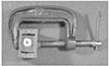
The muffle system introduced by Bramante5) and modified by McCann et al.6) was used to evaluate the root canal preparation. Teeth were mounted in the muffle with roots embedded in the acrylic resin. During root canal shaping procedure, two muffle halves were held together with a common C clamp (Figure 1).
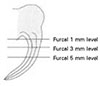
After sectioning the embedded roots horizontally at 1, 3, and 5 mm levels from the furcation with a microtome (Isomet™, Buehler Co., Lake Bluff, IL, U.S.A., Figure 2), teeth were remounted in the muffle system.
Root canal preparation
Forty roots were divided into four groups according to the rotary nickel-titanium instruments used: ProFile®, GT™ Rotary (Dentsply-Maillefer, Ballaigues, Switzerland), Quantec (Analytic Endodontics, Glendora, CA, U.S.A.), and ProTaper™ (Dentsply-Maillefer). Both the mesiobuccal and mesiolingual canals were instrumented using the crown-down technique with one of the above rotary nickel-titanium instruments. In each root, one canal was prepared with a straight up-and-down motion and the other canal was with anticurvature motion. Canals were instrumented until apical diameter had attained a size of 30. Each file was examined before and after use for any defects, and was wiped regularly to remove debris. Each instrument was used in only one canal before being replaced. Root canals were irrigated after each instrument use with 5 mL of water using a 27-gauge needle (Endo-Tips, Ultradent Products Inc., Utah, U.S.A.). RC-Prep™ (Stone Pharmaceuticals, Philadelphia, U.S.A.) was used as a lubricant. Canals were prepared with a crown-down technique according to the recommended sequences of the manufacturers by one operator who was a postgraduate student of Endodontic Program with one year of clinical experience of Ni-Ti rotary files. The operator practiced each instrumentation technique twenty times before experiment.
In the ProFile group, Orifice Shaper #3 and #2 were used until resistance was encountered (12 to 15 mm) for coronal shaping followed by 0.06/#25 and .06/#20 files to resistance (within 1 to 2 mm of the working length). For apical shaping, 0.04/#25 and 0.04/#30 files were used to the working length. In the GT Rotary group, 0.12/#20, 0.10/#20, 0.08/#20 and 0.06/#20 files were used sequentially until progression became difficult for coronal flaring. A 0.12/#20 file was used to a depth of 12 mm. 0.10/#20 to a depth of 14 mm, a 0.08/#20 to 16 mm and a 0.06/#20 to the working length. 0.04/#25 and 0.04/#30 files were used for apical shaping. In the Quantec group, a Quantec LX #1 (0.06/#25, 17 mm) was used to a depth of 11 to 12 mm for coronal flaring. For the deep canal shaping, 0.12/#25, 0.10/#25, 0.08/#25, and 0.06/#25 files were inserted in the canal as deep as possible. A 0.12/#25 file was used to 12 mm, a 0.10/#25 to 14 mm, a 0.08/#25 to 16 mm and finally 0.06/#25 and 0.05/#25 files were used to the working length. Apical shaping was done by an Accessory Quantec LX 0.02/#30 file. In the ProTaper group, a Shaping file No.1 (S1) was used first in the canal and moved apically to just short of the working length (16 mm). SX files were then used sequentially to resistance (13 to 14 mm) followed by S1 and S2 to working length for the shaping of the coronal two-thirds of the canal. Apical one-third was finished using F1, F2 and F3 sequentially to the working length with only one pecking motion for each instrument.
Assessment of shaping ability
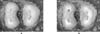
Shaping ability of each instrument was assessed by the evaluation of the root dentin thickness change. Pre- and post-instrumentation canal images were observed under a stereomicroscope (SZ40, Olympus Optical Co., Ltd., Tokyo, Japan), and stored in a computer using a CCD camera (GP-KR222, Panasonic, Osaka, Japan) and a commercial digitizing image program (micro VIDEO Studio 200 program, Pinnacle system, Brauschweig, Germany, Figure 3). The pre- and post-instrumentation canal images were superimposed and changes in root thickness were measured at distal side (danger zone) and mesial side (safe zone) of the canal using a computer program (Auto®CAD 2000, Autodesk Corp., San Rafael. CA, U.S.A.).
III. RESULTS
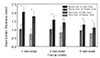
Root dentin thicknesses and its changes, in general, are shown in Figure 4. Before instrumentation, root dentin thickness at danger zone was significantly thinner than that at safe zone at each furcal level of 1, 3, and 5 mm (n = 80, p < 0.05). Regardless of the instruments and instrumentation techniques, root dentin thickness at danger zone remained significantly thinner than that at safe zone after instrumentation at all furcal levels (n = 80, p < 0.05). However, there was a tendency to remove more dentin at safe zone than at danger zone at 1 mm level compared to 3 and 5 mm levels without statistical significance.
In all instrument groups, there was no significant difference in the change of root dentin thickness between the straight up-and-down and the anticurvature motions at both danger and safe zones (p > 0.05, Tables 1 and 2).
Among instrumentation groups, ProTaper group removed significantly more dentin than in another groups at the furcal 3 mm level of the danger zone and at 1 and 3 mm levels of safe zone (p < 0.05, Table 1).
No perforations were noted in the study.
IV. DISCUSSION
Cross sections through the coronal third of furcated roots reveal that canals are not typically centered anatomically within their roots. Instead, they are often skewed toward the furcal-side concavities7). The mesial root of mandibular first molars was reported to have a distal surface concavity with a root thickness in this area of about 0.7 mm8), and is subject to perforation. In the present study, cross-section of the mesial roots of mandibular molars showed that root dentin thickness at distal side (danger zone) was significantly thinner than that at mesial side (safe zone) at all levels from the furcation too. This finding was in agreement with the previous reports7,8). Because root curvature is toward the distal side in general, this distal side is especially subject to strip perforation.
It was suggested to limit the circumferential filing to the areas of greatest bulk to prevent stripping perforation on the furcal side of the root canal4). Coronal flaring can reduce these undesirable aberrations, and it has also been recommended that they should be used like files, with an anticurvature motion toward the safe zone of the tooth9).
The nickel-titanium files were found to have two to three times more elastic flexibility of the stainless steel files in bending and torsion, as well as superior resistance to fracture10). Because of its high flexibility, the load on the cutting blade is greatly reduced in curved canals, which reduces stress on the instrument and the possibility of fracture. Several studies have reported that canals prepared with rotary nickel-titanium files of high taper were excellently tapered and that the use of these files reduced the incidence of canal aberration11-16) and have a decreased tendency for canal transportation and therefore remain better centered17).
It was suggested that the coronal two-thirds of the canal can easily be moved and relocated away from furcal danger and toward the greatest bulk of dentin when Gates Glidden burs are used in a step-back technique18). Gates Glidden burs were used to cut and remove dentin on just one or two of the outer walls of the canal and away from furcal danger.
Isom et al.18) compared root thickness in the mesial canals of lower molars before and after flaring with Gates Glidden burs by using a muffle system. Gates Glidden burs were used with either a straight up-and-down motion or with an anticurvature motion. At a level 2 mm apical to the furcation, the anticurvature method removed more dentin than the straight up and down18). However, there was no significant difference in the change of root dentin thickness between the straight up-and-down and the anticurvature motions in the present study. The reason of the difference of the result between the stainless steel instrument and nickel-titanium ones may be the difference of their flexibility. As nickel-titanium files induce less transportation of the canal, it may have less effectiveness in anticurvature filing. One of the rotary nickel-titanium instruments, LightSpeed caused significantly less transportation in 60 mesial canals in mandibular molars than did stainless steel or nickel-titanium manual files19). Therefore, the anticurvature motion of nickel-titanium rotary instruments seems to be less effective to cut the dentin of safe zone than stainless steel ones do.
Shape of the prepared root canal may be influenced by the design and taper of instruments. In the present study, ProTaper group removed significantly more dentin than other groups, especially at furcal 3 mm level at danger zone. The taper of the ProTaper files is bigger than the other files at the same level of the root canal, which may result in greatest reduction in the thickness of root canal dentin. Yun and Kim20) studied shaping abilities of four nickel-titanium rotary instruments in canals in plastic blocks. The finding of the present study is in agreement with theirs in that ProTaper cut more dentin than any other instruments tested.
Muffle system was used in this study. The model system allowed direct comparison of the four instrumentation techniques at three canal levels. Although the technique described by Bramante et al.5) is an excellent method for the comparison of original and shaped canals, some problems21) were encountered. First, during sectioning, 0.4 mm over of root material was lost. The additional loss of root material was caused by the lateral movement of band saw. Secondly, not all sections were at right angles to the canal. In curved canals the loss of root material and some oblique-sectioned surfaces acted as ledges that hindered the passage of the file through the canal to the working length. In severely curved canals, this even prevented further instrumentation, irrespective of the technique used. Further investigations should try to minimize the width of dentine lost during root sectioning. This is best achieved by reducing the lateral movements of the band saw or by using a different sectioning technique.
Therefore, it was concluded that there was no difference between up-and-down motion and anticurvature one in removing root dentin with nickel-titanium rotary instruments in the condition of the present study, which indicates that anticurvature motion with nickel-titanium rotary instruments may not as effective as that with stainless steel ones. ProTaper removed more root dentin than GT Rotary, Quantec, and ProFile especially at furcal 3 mm level.
Further research is needed to evaluate influence of working length, apical diameter and canal curvature on the shaping ability of rotary nickel-titanium instruments to prepare root canal at danger zone.


 XML Download