

 PDF
PDF ePub
ePub Citation
 Print
Print


Abstract
Established microleakage tests have their own disadvantages. In this study, 3D reconstruction method was tried to overcome these disadvantages.
Four types of microleakage tests were used and relationships among them were estimated: penetrated dye volume; marginal adaptability; degree of dye penetration and relative penetrated length to cavity wall.
Twenty-four Class V cavities were bulk filled with composite (Esthet X) following surface treatments: N group (no treatment); E group (etching only); T group (etching + Prime & Bond NT). 50% silver nitrate was used as a dye solution after thermocycling (5℃ & 55℃, 1,000 times). Teeth were serially ground with a thickness of 0.2 mm. Volume of dye penetration was estimated from a three-dimensionally reconstructed image with a software (3D-DOCTOR). Percentage of margin without gap was estimated from SEM and degree of dye penetration and the relative length of dye penetration to overall cavity wall were also estimated.
ANOVA and Scheffe test for dye volume, Kruskal-Wallis and Mann-Whitney test for marginal quality, Spearman's rho test for checking of relationships among methods were used.
The results were as follows:
1. Dye penetration could be seen from several directions, furthermore, its volumetric estimation was possible.
2. Reverse relationship was found between dye volume and marginal quality (r = -0.881 / p = 0.004).
3. Very low relationship was seen between dye volume and two-dimensional tests (degree of dye penetration and relative length). However, 2D evaluation methods showed high relationship (p = 0.002-0.054) each other.
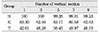
4. Three times vertical section could be recommended as a 2D test.
Figures and Tables
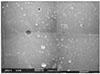 | Figure 1Marginal adaptation from SEM evaluation: Percentage of gap-free margin (A; red) to cavity perimeter (B; blue) (SEM image; × 35)
|
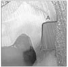 | Figure 4Evaluation criteria about percentage of dye penetration: Percentage of length of dye penetration (B; red) to overall cavity wall length (A; blue)
|
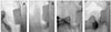 | Figure 5Evaluation criteria about degree of dye penetration: 0 - No dye penetration (A); 1 - Dye penetration less than half the length of the gingival or occlusal wall (B); 2 - Dye penetration up to the full length of the gingival or occlusal wall (C); 3 - Dye penetration along the axial wall (D)
|
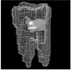
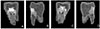 | Figure 6Three-dimensionally reconstructed tooth (group N): A; buccal view, B; mesial view, C; lingual view, D; distal view
|
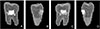 | Figure 7Three-dimensionally reconstructed tooth (group E): A; buccal view, B; mesial view, C; lingual view, D; distal view
|
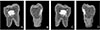 | Figure 8Three-dimensionally reconstructed tooth (group T): A; buccal view, B; mesial view, C; lingual view, D; distal view
|
References
1. Saboia Vde P, Pimenta LA, Ambrosano GM. Effect of collagen removal on microleakage of resin composite restorations. Oper Dent. 2002. 27(1):38–43.
2. Adamo HL, Buruiana R, Schertzer L, Boylan RJ. A comparison of MTA, Super-EBA, composite and amalgam as root end filling materials using a bacterial microleakage model. Int Endod J. 1999. 32:197–203.

3. Hembree JH Jr, Andrew JT. Microleakage of several class V anterior restorative materials: a laboratory study. J Am Dent Assoc. 1978. 97(2):179–183.
4. Manhart J, Chen HY, Mehl A, Weber K, Hickel R. Marginal quality and microleakage of adhesive class V restorations. J Dent. 2001. 29(2):123–130.
5. Youngson CC, Jones JC, Fox K, Smith IS, Wood DJ, Gale M. A fluid filtration and clearing technique to assess microleakage associated with three dentine bonding systems. J Dent. 1999. 27(3):223–233.
6. Von Fraunhofer JA, Adachi EI, Barnes DM, Romberg E. The effect of tooth preparation on microleakage behavior. Oper Dent. 2000. 25(6):526–533.
7. Iwami Y, Yamamoto H, Ebisu S. A new electrical method for detecting marginal leakage of in vitro resin restorations. J Dent. 2000. 28(4):241–247.
8. Uno S, Finger WJ, Fritz UB. Effect of cavity design on microleakage of resin-modified glass ionomer restorations. Am J Dent. 1997. 10(1):32–35.
9. Estafan D, Pines MS, Erakin C, Fuerst PF. Microleakage of Class V restorations using two different compomer systems: an in vitro study. J Clin Dent. 1999. 10(4):124–126.
10. Pilo R, Ben-Amar A. Comparison of microleakage for three one-bottle and three multiple-step dentin bonding agents. J Prosthet Dent. 1999. 82(2):209–213.
11. Ziskind D, Avivi-Arber L, Haramati O, Hirschfeld Z. Amalgam alternatives-microleakage evaluation of clinical procedures. Part I: direct composite/composite inlay/ceramic inlay. J Oral Rehabil. 1998. 25(6):443–447.
12. Baldissara P, Comin G, Martone F, Scotti R. Comparative study of the marginal microleakage of six cements in fixed provisional crowns. J Prosthet Dent. 1998. 80(4):417–422.
13. Hatibovic-Kofman S, Wright GZ, Braverman I. Microleakage of sealants after conventional, bur, and air-abrasion preparation of pits and fissures. Pediatr Dent. 1998. 20(3):173–176.
14. Kazemi RB, Spangberg LS. Effect of reduced air pressure on dye penetration in standardized voids. Oral Surg Oral Med Oral Pathol Oral Radiol Endod. 1995. 80(6):720–725.
15. Grande RH, Ballester R, Singer Jda M, Santos JF. Microleakage of a universal adhesive used as a fissure sealant. Am J Dent. 1998. 11(3):109–113.
16. Wibowo G, Stockton L. Microleakage of class II composite restorations. Am J Dent. 2001. 14(3):177–185.
17. Arnold WH, Gaengler P, Kalkutschke L. Three-dimensional reconstruction of approximal subsurface caries lesions in deciduous molars. Clin Oral Investig. 1998. 2(4):174–179.
18. Mikrogeorgis G, Lyroudia KL, Nikopoulos N, Pitas I, Molyvdas I, Lambrianidis TH. 3D computer-aided reconstruction of six teeth with morphological abnormalities. Int Endod J. 1999. 32(2):88–93.
19. Jacobs R, Adriansens A, Verstreken K, Suetens P, van Steenberghe D. Predictability of a three-dimensional planning system for oral implant surgery. Dentomaxillofac Radiol. 1999. 28(2):105–111.
20. Jung EH, Shin DH. Morphological analysis of C-shaped root using 3D reconstruction. J Korean Acad Conserv Dent. 2002. 27(4):421–431.
21. Veis A, Lambrianides T, Nicolaou A. Area-metric analysis of dye leakage for evaluation of sealing ability of root canal obturation techniques. Endod Dent Traumatol. 1996. 12(5):222–226.
22. Gale MS, Darvell BW, Cheung GS. Three-dimensional reconstruction of microleakage pattern using a sequential grinding technique. J Dent. 1994. 22(6):370–375.
23. Pashley DH, Depew DD. Effects of the smear layer, Copalite and oxalate on microleakage. Oper Dent. 1986. 11:95–102.
24. Bullard RH, Leinfelder KF, Russell CM. Effect of coefficient of thermal expansion on microleakage. J Am Dent Assoc. 1988. 116:871–874.
25. Heys RJ, Fitzgerald M. Microleakage of three cement bases. J Dent Res. 1991. 70:55–58.
26. Pachuta SM, Meiers JC. Dentin surface treatment and glass ionomer microleakage. Am J Dent. 1995. 8:187–190.
27. Doerr CL, Hilton TJ, Hermesch CB. Effect of thermocycling on the microleakage of conventional and resin-modified glass ionomer. Am J Dent. 1996. 9:19–21.
28. Tate WH, Friedl KH, Power JM. Bond strength of composites to hybrid ionomers. Oper Dent. 1996. 21:147–152.
29. Youngson CC, Jones JC, Manogue M, Smith IS. In vitro dentinal penetration by tracers used in microleakage studies. Int Endod J. 1998. 31(2):90–99.
30. Hakimeh S, Vaidyanathan J, Houpt ML, Vaidyanathan TK, Von Hagen S. Microleakage of compomer class V restorations: effect of load cycling, thermal cycling, and cavity shape differences. J Prosthet Dent. 2000. 83(2):194–203.
31. Gladys S, van Meerbeek B, Lambrechts P, Vanherle G. Microleakage of adhesive restorative materials. Am J Dent. 2001. 14(3):170–176.
32. Van Meerbeek B, Perdigão J, Gladys S, Lambrechts P, Vanherle G. Schwartz R, Summitt JB, Robbins JW, editors. Enamel and dentin adhesion. Fundamentals of operative dentistry. A contemporary approach. 1996. Chicago, IL: Quintessence Publishing Co., Inc;141–186.
33. Crim GA. Marginal leakage of visible light-cured glass ionomer restorative materials. J Prosthet Dent. 1993. 69:561–563.
34. Hilton TJ, Ferracane JL. Cavity preparation factors and microleakage of Class II composite restorations filled at intraoral temperatures. Am J Dent. 1999. 12:123–130.
35. Feilzer AJ, de Gee AJ, Davidson CL. Setting stress in composite resin in relation to the configuration of the restoration. J Dent Res. 1987. 66:1636–1639.
36. Feilzer AJ, de Gee AJ, Davidson CL. Curing contraction of composites and glass-ionomer cements. J Prosthet Dent. 1988. 59:297–300.
37. Carvalho RM, Pereira JC, Yoshiyama M, Pashley DH. The influence of stress development versus stress relief. Oper Dent. 1996. 21:17–24.
38. Asmussen E, Munksgaard EC. Status of dentin adhesives and impact on cavity design and filling techniques. Int Dent J. 1988. 38:97–104.
39. Santini A, Mitchell S. Microleakage of composite restorations bonded with three new dentin bonding agents. J Esthet Dent. 1998. 10(6):296–304.
40. Mixson J, Eick JD, Chappell RP, Tira DE, Moore DL. Comparison of two-surface and multi-surface scoring methodologies for in vitro microleakage studies. Dent Mater. 1991. 7(3):191–196.
 XML Download