

 PDF
PDF ePub
ePub Citation
 Print
Print


I. Introduction
Apicoectomy with retrograde filling is a well-established technique to treat persistent periapical infections in which conventional root treatment is not accessible. Retrofilling is the procedure by which the apex of the root is resected after its surgical exposure and an inert nontoxic material is packed into a prepared cavity at the apical end.
Various materials have been advocated as retrofillings, but non have met all of the criteria set up in the literature1). Retrofilling materials should be biocompatible with apical tissue, dimensionally stable, nonabsorbable, radiopaque, bacteriostatic, and capable of providing a tight apical seal2,3).
It has been documented that Super-EBA provide clinical success rates 95%, respectively, over a 6-month to 10-year observation period4). Super-EBA cement provides an optimum seal and minimal tissue toxicity5).
Torabinejad et al.6,16) introduced MTA as a root end filling material and showed that MTA leaked less than Super-EBA or amalgam even in presence of blood. But super-EBA cement and MTA are difficult to mix and handle requiring more effort and practice than other root-end filling materials.
The frequency of healing after retrograde root-end cover performed with a resin composite (Retroplast) and the dentin-bonding agent Gluma (Bayer AG, Gluma 1 and 2) has previously been reported7).
Greer et al.8) showed that Dyract and Geristore are equal or superior to IRM and equvialent to Super-EBA in their ability to reduce apical leakage when used as retrofilling materials.
There is few studies to compare the apical leakages obtained with super-EBA, MTA, and Dyract flow as a retrograde filling matierial.
The purpose of this study was to compare the apical sealing ability of Super-EBA cement (Super-EBA™, Bosworth Company, Illinois, USA), MTA(ProRoot™, Dentsply, Tulsa Dental, Maillefer; Oklahoma, USA) and Dyract-flow (Dentsply/Detrey, Konstantz, Germany) as retrofilling materials.
II. Materials and Methods
Forty-eight extracted human teeth, with fully developed apices, straight and single root canals were used in this study. The teeth were stored in 0.9% isotonic saline at room temperature at all times. Specimens were soaked in 5.25% NaOCl for 30 minutes, and the remaining periodontal tissue and calculus were removed.
To facilitate instrumentation, the crown portion of each tooth was removed using a high speed fissure bur under water spray. Only teeth with roots 12 to 15 mm long were used.
Working length was determined by placing a #10 k flie (k-flexofile, Dentsply Maillefer ; Ballaigues, Switzerland) until it was just seen penetrating the foramen, after which 1mm was subtracted from this and the length was recorded. The root canals were prepared to a #40 apical canal size with Profiles (Profile .06, Dentsply Maillefer, Ballaigues, Switzerland) by crown down pressureless technique. The teeth were dried thoroughly with paper points and obturated with gutter-percha using a continuous wave of condensation technique.
Apicoectomies were performed by sectioning the apical 3mm using a high speed fissure bur under water spray. A root end cavities were prepared to a depth of 3mm using an ultrasonic device (Kis™ Microsurgical instruments, Swiss Machining Inc., San Diego, USA).
The teeth were randomly divided into three experimental groups of 14 teeth each and two control groups of two teeth each as follows.
Group 1 (14 teeth): Super-EBA was mixed according to manufacturer's specifications and placed into the root-end preparation with a plastic instrument and tamped down with a miniature condenser, then allowed to set.
Group 2 (14 teeth): MTA was mixed with sterile distilled water and placed into the root-end preparations with a plastic instrument, tamped down with a miniature condenser and stored at 100% humidity.
Group 3 (14 teeth): Dyract-flow was placed into the root-end preparation after applying Prime & Bond NT (Dentsply/Detrey, Konatantz, Germany) and curing without etching, then cured for 40 seconds.
Group 4 (2 teeth): Apical cavities were left unfilled. These roots were used as positive controls to prove that the electrolyte can penetrate to the full length of the root canal.
Group 5 (2 teeth): The apical cut surface of these roots was painted completely with a double layer of nail varnish to seal the apical opening of the canals. These roots were used as negative controls to prove that the electrolyte can be prevented from apical penetration.
The obturated gutter-percha was then removed from each canal as completely as possible by using System B Heatsource (Model 1005, Analytic Technology, Redmond, WA, USA), except the negative controls. The entire root surface of each tooth except the cutting surface of the apical end, was sealed with two coats of nail varnish.
Copper wires that were used as experimental electrodes were inserted into the canals in contact with the root end filling materials. Approximately 0.5mm of insulation coat was stripped off at both ends of the wire. A small piece of sticky wax was adapted around the wire at the occlusal opening to stabilize the wire.
All the specimens were placed in the bottles. The copper wire as an experimental electrode was fixed in the canal and extended to the outside of the bottle cap. The stainless-steel wire that acted as a standard electrode was located at the center of the bottle. A 0.9% NaCl solution as an electrolyte was placed in the bottle until the apical half of the roots were covered.
The electrical resistance between the standard and experimental electrodes was measured over a period of four weeks using a multitester (Dagatron 2021, Dagatronics Corp., Seoul, Korea).
The data were statistically analyzed using Repeated measures of ANOVA.
III. Results
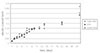
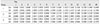
Increasing leakage with time was observed in all experimental groups. The leakage increased markedly within the first four days (p < 0.0001). Positive controls (group 4) showed immediate leakage with which increased to entire leakage with complete penetration of the electrolyte solution whereas the negative control (group 5) exhibited no leakage.
IV. Discussion
It has been documented that Super-EBA provide clinical success rates 95%, respectively, over a 6-month to 10-year observation period4). Super-EBA cement provides an optimum seal and minimal tissue toxicity5). But, Super-EBA is difficult to mix, requring more effort and practice than most other root-end filling materials.
MTA has been reported equal or superior to Super-EBA with respect to dye and bacterial leakage and marginal adaptation6,9-13). Also, formation of cementum and periodontal ligament fibers has been shown adjacent to MTA14,15). Bleeding in the field can interfere with MTA placement, although the final set may not be adversely affected by blood contamination16). In spite of these good properties, MTA is very expensive and handling of MTA is difficult.
The results of this study show that sealing ability of Dyract-flow appeared comparable with Super-EBA and MTA in terms of working time, ease of manipulation and handling. Dyract-flow is a compomer, which is a mixture of composite resin and glass ionomer cement. This material is becoming popular in restorative dentistry, because this possesses the strength and esthetic quality of the composite restorative material, while bonding without need for acid-etching to dentin. This hybrid is easier to place than the older materials due to their one-component, no-mix direct delivery system17). Moreover, an in vivo intraosseous implantation study in the rabbit showed that a compomer, Dyract, has as good biocompatibility as Super EBA. The thickness of the fibrous interposition between the materials and the new-formed bone was more frequently seen with Super EBA and area of direct contact between bone and Dyract was formed more frequently than Super EBA18). Significantly, they exhibit biocompatability in that gingival tissues appear to adhere to them, resulting in a postoperative gain in clinical attachment when placed subgingivally19). This feature may allow fibroblasts of the periodontal ligament to attach to and reform around a root apex in which a compomer root-end filling is placed.
On the other hand, the inconvenience of use of Dyract include the need for a bonding agent and the application of which increases preparation time. Moreover, despite its improved properties, Dyract-flow, while in an unset state, is probably much more sensitive to moisture than Super-EBA and MTA. Whether its physical and biological properties are altered if contaminated by blood is still to be investigated.
Apical seal was evaluated indirectly by assessing marginal adaptation and directly by observing leakage of various tracers into retrofilled root canals.
Jacobson and VonFrauhofer20) described an electrochemical method for microleakage measurement. Delivanis and Chapman21) found this method to be effective and reliable. They considered it as the only method by which rapid quantitative results can be obtained.
Different methods have been used in the study of marginal leakage. Despite the wide use of dye penetration and autoradiography for evaluation of marginal leakage, Delvanis and Champman21) found them to have significant limitations and a significant margin of error. If these methods are not precisely controlled, variables can potentially change the results. They found the electrochemical technique to be effective and reliable. They described that electrochemical method could provide quantitative measurements of apical leakage and the opportunity to study leakage over a continuous time period. The time elapsed between immersion and current flow reflects the penetration of electrolyte and the magnitude of the current indicates the degree of leakage. This is because the current magnitude is controlled by diffusion of electroactive ions to the electrode surface and is directly proportional to the surface area of the electrode. In this study, the magnitude of the current was measured as the electric resistance between standard and experimental electrodes.
V. Conclusions
The purpose of this study was to assess and compare the apical seal obtained with Super-EBA, MTA and Dyract-flow when used as retrofilling materials. Leakage was measured using an electrochemical technique for 4 weeks.
According to this study, the results were as follows.
The results of this study suggest that the sealing ability of Dyract-flow is equal to the Super-EBA and MTA, and that Dyract-flow may be an alternative to other materials for root-end filling.
 XML Download