

 PDF
PDF ePub
ePub Citation
 Print
Print


Abstract
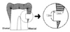
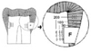
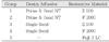
The purpose of this study was to compare the microhardness and the fluoride content of enamel and dentin around fluoride- or non fluoride-containing restorations. Forty extracted human teeth were used and prepared cervical cavities on proximal surface. Experimental teeth were divided into five groups. Group 1 : Prime & Bond NT and Z100, Group 2 : Prime & Bond NT and F2000, Group 3 : Scotchbond Multi-Purpose and Z100, Group 4 : Scothcbond Multi-purpose and F2000, Group 5 : Fuji II LC. The cavities were filled with dentin adhesives and restorative materials. After each tooth was bisected, one half was tested microhardness and the other half was analyzed the fluoride at the enamel and dentin by an EPMA-WDX device. The results were as follows:
1. There was no statistical difference among the microhardness of enamel surface in all group.
2. The microhardness at dentin of 100 µm point in Group 2 and 20 µm point in Group 4 was lower than that of normal dentin (p>0.05).
3. There was no statistical difference among the fluoride content of enamel surface in all group.
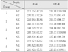
4. The fluoride content at the dentin of 30 µm point in Group 2 and 5 were higher than those at 100 µm and 200 µm point in Group 2 and normal dentin (p<0.05).
5. At the dentin of 30 µm point, Group 2 showed higher fluoride content than Group 1 and 3, and Group 5 showed higher fluoride content than other groups.
Figures and Tables


References
1. MacInnis WA, Ismail A, Brogan H. Placement and replacement of restorations in a military population. J Can Dent Assoc. 1991. 57:227–231.
2. Corpron RE, More FG, Clark JW, Korytnicki D, Kowalski CJ. In vivo remineralization of artificial enamel lesions by a fluoride dentifrice or mouth rinse. Caries Res. 1986. 20:48–55.

3. Norman RD, Mehra RV, Swartz ML, Philips RW. Effects of restorative materials on plaque composition. J Dent Res. 1972. 51:1596–1601.
4. Donly KJ, Segura A, Wefel JS, Hogan MM. Evaluating the effects of fluoride-releasing dental materials on adjacent interproximal caries. J Am Dent Assoc. 1999. 130:817–825.
5. Hicks MJ, Flaitz CM. Resin-modified glass ionomer restorations and in vitro secondary caries formation in coronal enamel. Quintessence Int. 2000. 31:570–578.
6. Retief DH, Bradley EL, Denton JC, Switzer P. Enamel and cementum fluoride uptake from a glass ionomer cement. Caries Res. 1984. 18:250–257.
7. Torii Y, Itota T, Okamoto M, Nakabo S, Nagaminie M, Inoue K. Inhibition of artificial secondary caries in root by fluoride-releasing restorative materials. Oper Dent. 2001. 26:36–43.
8. Diaz-Arnold AM, Holmes DC, Wistrom DW, Swift EJ Jr. Short-term fluoride release/uptake of glass ionomer restoratives. Dent Mater. 1995. 11:96–101.
9. Dunne SM, Goolnik JS, Millar BJ, Seddon RP. Caries inhibition by a resin-modified and a conventional glass ionomer cement in vitro. J Dent. 1996. 24:91–94.
10. Takahashi K, Emilson CG, Birkhed D. Fluoride release in vitro from various glass ionomer cements and resin composites after exposure to NaF solutions. Dent Mater. 1993. 9:350–354.
11. Eliades G, Kakaboura A, Palaghias G. Acid-base reaction and fluoride release profiles in visible light-cured polyacid modified composite restoratives. Dent Mater. 1998. 14:57–63.
12. Forsten L. Fluoride release and uptake by glassionomers and related materials and its clinical effect. Biomaterials. 1998. 19:503–508.
13. Verbeeck RM, De Maeyer AP, Marks LA, De Moor JG, De Witte AM, Trimpeneers LM. Fluoride release process of (resin-modified) glass-ionomer cements versus (polyacid-modified) composite resins. Biomaterials. 1998. 19:509–519.
14. Yap AUJ, Khor E, Foo SH. Fluoride release and antibacterial properties of new-generation tooth-colored restoratives. Oper Dent. 1999. 24:297–305.
15. Momoi Y, McCabe JF. Fluoride release from light-activated glass-ionomer restorative cements. Dent Mater. 1993. 9:151–154.
16. Swartz ML, Phillips RW, Clark HE. Long-term F release from glass-ionomer cements. J Dent Res. 1984. 63:158–160.
17. Yap AUJ, Tham SY, Zhu LY, Lee HK. Short-Term Fluoride release from various aesthetic restorative materials. Oper Dent. 2002. 27:259–265.
18. Araujo FB, Godoy FG, Cury JA, Conceicao EN. Fluoride release from fluoride-containing materials. Oper Dent. 1996. 21:185–190.
19. Itota T, Okamoto M, Sato K, Nakabo S, Nagamine M, Torii Y, Inoue K. Release and recharge of fluoride by restorative materials. Dent Mater J. 1999. 18:347–353.
20. Preston AJ, Mair LH, Agalamanyi EA, Higham SM. Fluoride release from aesthetic dental materials. J Oral Rehabil. 1999. 26:123–129.
21. Helvatjoglu-Antoniades M, Karantakis P, Papadogiannis Y, Kapetanios H. Fluoride release from restorative materials and a luting cement. J Prosthet Dent. 2001. 86:156–164.
22. Francci C, Deaton TG, Arnold RR, Swift EJ Jr. Fluoride release from restorative materials and its effects on dentin demineralization. J Dent Res. 1999. 78:1647–1654.
23. Duckworth RM, Lynch RJM. Fluoride uptake to demineralised enamel: A comparison of sampling techniques. Caries Res. 1998. 32:417–421.
24. Jones FH, Hutton BM, Hadley PC, Eccles AJ, Steele TA, Billington RW, Pearson GJ. Fluoride uptake by glass ionomer cements: a surface analysis approach. Biomaterials. 2003. 24:107–119.
25. Kawai K, Tantbirojin D, Kamalawat AS, Hasegawa T, Retief DH. In vitro enamel and cementum fluoride uptake from three fluoride-containing composites. Caries Res. 1998. 32:463–469.
26. Raven SJ, Schafer F, Duckworth RM, Gilbert RJ, Parr TA. Comparison between evaluation methods for the anti-caries efficacy of monofluorophosphate-containing dentifrices. Caries Res. 1991. 25:130–137.
27. Papagiannoulis L, Kakaboura A, Eliades G. In vivo vs in vitro anticariogenic behavior of glass-ionomer and resin composite restorative materials. Dent Mater. 2002. 18:561–569.
28. Pereira PNR, Inokoshi S, Tagami J. In vitro secondary caries inhibition around fluoride releasing materials. J Dent. 1998. 26:505–510.
29. Yamamoto H, Iwami Y, Unezaki T, Tomii Y, Ebisu S. Fluoride uptake in human teeth from fluoride-releasing restorative material in vivo and in vitro: Two-dimensional mapping by EPMA-WDX. Caries Res. 2001. 35:111–115.
30. Hotta M, Li Y, Sekine I. Mineralization in bovine dentin adjacent to glass-ionomer restorations. J Dent. 2001. 29:211–215.
31. Reintsema H, Arends J. An in vivo study of microhardness and fluoride uptake in partially demineralized human enamel covered by plaque. J Dent Res. 1988. 67:471–473.
32. Samuel SM, Rubinstein C. Microhardness of enamel restored with fluoride and non-fluoride releasing dental materials. Braz Dent J. 2001. 12:35–38.
33. Nakabayashi N, Nakamura M, Yasuda N. Hybrid layer as a dentin bonding mechanism. J Esthet Dent. 1991. 3:133–138.
34. Benelli EM, Serra MC, Rodrigues AL Jr, Cury JA. In situ anticariogenic potential of glass ionomer cement. Caries Res. 1993. 27:280–284.
35. Dijkman GE, de Vries J, Arends J. Secondary caries in dentine around composites: a wavelength-independent microradiographical study. Caries Res. 1994. 28:87–93.
36. Dionysopoulos P, Kotsanos N, Pagadogiannis Y, Konstantinidis A. Artificial secondary caries around two new F-containing restoratives. Oper Dent. 1998. 23:81–86.
37. Guha-Chowdhury N, Clark AG, Sissons CH. Inhibition of purified enolases from oral bacteria by fluoride. Oral Microbiol Immunol. 1997. 12:91–97.
38. Marquis RE. Antimicrobial actions of fluoride for oral bacteria. Can J Microbiol. 1995. 41:955–964.
39. Dijkman GE, Arends J. Secondary caries in situ around fluoride-releasing light-curing composites: A quantitative model investigation on four materials with a fluoride content between 0 and 26 vol%. Caries Res. 1992. 26:351–357.
40. Corpron RE, More FG, Mount G. Comparison of fluoride profiles by SIMS with mineral density of subsurface enamel lesions treated intra-orally with a fluoride-releasing device. J Dent Res. 1992. 71:828–831.
41. Kotsanos N. An intraoral study of caries induced on enamel in contact with fluoride-releasing restorative materials. Caries Res. 2001. 35:200–204.
42. Han L, Abu-Bakr N, Okamoto A, Iwaku M. Study of the fluoridated adhesive resin cement--fluoride release, fluoride uptake and acid resistance of tooth structures. Dent Mater J. 2001. 20:114–122.
43. Boyde A, et al. Application of the scanning electron probe X-ray microanalyzer to dental tissues. J Ultrastruct Res. 1961. 5:201–207.
44. Feilzer AJ, De Gee AJ, Davidson CL. Relaxation of polymerization contraction shear stress by hygroscopic expansion. J Dent Res. 1990. 69:36–39.
45. Mazzaoui SA, Burrow MF, Tyas MJ. Fluoride release from glass ionomer cements and resin composites coated with a dentin adhesive. Dent Mater. 2000. 16:166–171.
 XML Download