

 PDF
PDF ePub
ePub Citation
 Print
Print


Abstract
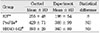
The purpose of this in vitro study was to evaluate the effect of surface defects and cross-sectional configuration of NiTi rotary files on the fatigue life under cyclic loading. Three NiTi rotary files (K3™, ProFile®, and HERO 642®) with #30/.04 taper were evaluated. Each rotary file was divided into 2 subgroups: control (no surface defects) and experimental group (artificial surface defects). A total of six groups of each 10 were tested. The NiTi rotary files were rotated at 300rpm using the apparatus which simulated curved canal (40 degree of curvature) until they fracture. The number of cycles to fracture was calculated and the fractured surfaces were observed with a scanning electron microscope. The data were analyzed statistically. The results showed that experimental groups with surface defects had lower number of cycles to fracture than control group but there was only a statistical significance between control and experimental group in the K3™ (p<0.05). There was no strong correlation between the cross-sectional configuration area and fracture resistance under experimental conditions. Several of fractured files demonstrated characteristic patterns of brittle fracture consistent with the propagation of pre-existing cracks.
This data indicate that surface defects of NiTi rotary files may significantly decrease fatigue life and it may be one possible factor for early fracture of NiTi rotary files in clinical practice.
Figures and Tables
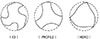
Figure 2
The apparatus developed for fracturing NiTi rotary files under cyclic loading (Schneider's curvature: 40°).
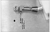
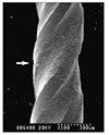
Figure 4
A SEM image of the fractured surface of HERO 642® file after cyclic loading: a control group (×150).
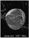
Figure 5
A higher magnification view of the rectangular area shown in Figure 4. Ultimate ductile fracture region that are typically characterized with microvoid formation and dimpling are seen (×1000).
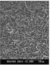
Figure 6
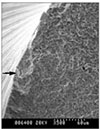
SEM image of the fractured surface of ProFile® after cyclic loading (experimental group, ×150): The left square area shows a region of brittle fracture (BF) which transit to ultimate ductile failure (DF).
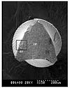
Figure 7
A higher magnification view of brittle fracture (BF) area shown in Figure 6. A region of brittle fracture is shown originating from the pre-existing surface defects (×500).

References
1. Mize SB, Pruett JP, Clement DJ, Carnes DL. Effect to sterilization on cyclic fatigue of rotary nickel-titanium endodontic instruments. J Endod. 1998. 24(12):843–847.

2. Pruett JP, Clement DJ, Carnes DL. Cyclic fatigue testing of nickel-titanium endodontic instruments. J Endod. 1997. 23(2):77–85.
3. Haikel Y, Serfaty R, Bateman G, Senger B, Allemann C. Dynamic and cyclic fatigue of engine-driven rotary nickel-titanium endodontic instruments. J Endod. 1999. 25(6):434–440.
4. Kuhn G, Tavernier B, Jordan L. Influence of structure on Nickel-Titanium endodontic instrument failure. J Endod. 2001. 27(8):516–520.
5. Karn T. Fractographic analysis of experimentally separated NiTi rotary files. 2003. University of Connecticut;MS Thesis.
6. Li UM, Lee BS, Shih CT, Lan WH, Lin CP. Cyclic fatigue of endodontic nickel-titanium rotary instruments: static and dynamic tests. J Endod. 2002. 28(6):448–451.
7. Turpin YL, Chagneau F, Vulcain JM. Impact of two theoretical cross-sections on torsional and bending stresses of nickel-titanium root canal instrument models. J Endod. 2000. 26(7):414–417.
8. Jeon IS, Spangberg LS, Yoon TC, Kazemi RB, Kum KY. Smear layer production of three NiTi rotary reamers in straight root canals. A SEM study. Oral Surg Oral Med Oral Pathol Oral Radiol Endod. 2003. 96(5):601–607.
 XML Download