

 PDF
PDF ePub
ePub Citation
 Print
Print


Abstract
Objectives
The purpose of this study was to determine whether pH and time has any influence on the degradation behavior of composite restoration by analyzing the leached monomers of dental composites qualitatively and quantitatively after storage in acetate buffer solution as a function of time using high performance liquid chromatography (HPLC) / mass spectrometer.
Materials and Methods
Three commercial composite restorative resin materials (Z-250, Heliomolar and Aeliteflo) with different matrix structure and filler composition were studied. Thirty specimens (7mm diameter×2mm thick) of each material were prepared. The cured materials were stored in acetate buffer solution at different pH (4, 7) for 1, 7 and 45days. As a reference, samples of unpolymerized composite materials of each product were treated with methanol (10 mg/ml). Identification of the various compounds was achieved by comparison of their mass spectra with those of reference compound, with literature data, and by their fragmentation patterns. Data were analysed statistically using ANOVA and Duncan's test.
Results
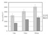
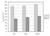
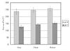
1. Amounts of leached TEGDMA in Aeliteflo were significantly larger than those of UDMA in Z-250 and Heliomolar at experimental conditions of different storage time and pH variation (p < 0.001).
2. As to comparison of the amounts of leached monomers per sorage time, amounts of leached TEGDMA in Aeliteflo and UDMA in Z-250 and Heliomolar were increased in the pH 4 solution more significantly than in the pH 7 solution after 1day, 7days and 45days, respectively (p < 0.001).
3. In total amounts of all the leached monomers with storage times, the overall amounts of pH 4 extracts were larger than those of pH 7 extracts for all resin groups, but there was no significant difference (p > 0.05).
Figures and Tables
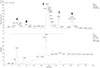
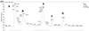
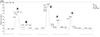
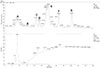
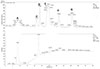
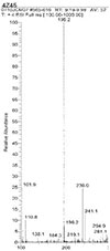
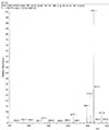
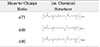
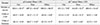
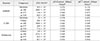
Figure 9
LC / MS - chromatogram of pH 7 extract from Heliomolar (Polymerized material)
Peak A ; Internal caffeine standard, fragmented methyl methacrylate, methacrylic acid, etc.
Peak B ; TEGDMA (triethyleneglycol dimethacrylate)
Peak C ; UDMA (urethane dimethacrylate)
Peak D ; Bis-GMA (Bisphenol A diglycidyl ether dimethacrylate)
Peak E ; Unidentified, probably related to Bis-EMA (Ethoxylated bisphenol A dimethacrylate)
Peak F,G ; A certain dimer or oligomer


References
1. Antonucci JM, Bowen RL. Dimethacrylates derived from hydroxylbenzoic acid. J Dent Res. 1976. 55:8–15.
2. Asmussen E. Factors affecting the quantity of remaining double bonds in restorative resin polymers. Scand J Dent Res. 1982. 90:490–496.

3. Antonucci JM, Toth EE. Extent of polymerization of dental resins by differential scanning calorimetry. J Dent Res. 1983. 62(2):121–125.
4. Ruyter IE, Oysaed H. Compressive creep of light cure resin based restorative materials. Acta Odontol Scand. 1982. 40:319–324.
5. Hanks CT, Craig RG, Diehl ML, Pashley DH. Cytotoxity of dental composites and other materials in a new in vitro device. J Oral Pathol. 1988. 17:396–403.
6. Ferracane JL, Berge HX, Condon JR. In vitro aging of dental composites in water-effect of degree of conversion, filler volume, and filler/matrix coupling. J Biomed Mater Res. 1998. 42:465–472.
7. Ferracane JL, DeWald JP. A comparison of four modes of evaluating depth of cure of light activated composite. J Dent Res. 1987. 66(3):727–730.
8. Ferracane JL, Condon JR. Rate of elution of leachable components from composite. Dent Mater. 1990. 6:282–287.
9. Inoue K, Hayashi I. Residual monomer (Bis-GMA) of composite resins. J Oral Rehabil. 1982. 9:493–497.
10. Tanaka K, Taira M, Shintani H, Wakasa K. Residual monomer(TEGDMA and Bis-GMA)of a set visable-light-cured dental composite resin when immersed in water. J Oral Rehabil. 1991. 18:353–362.
11. Dickens SH, Stansbury JW, Floyd CJ. Effects of chemical composition on cure properties of dental resins. J Dent Res. 1999. 78:1459–1463.
12. Ferracane JL. Elution of leachable components from composites. J Oral Rehabil. 1994. 21:441–452.
13. Gerzina TM, Hume WR. Effect of Dentin on the release of TEGDMA from resin composite in vitro. J Oral Rehabil. 1994. 21:463–468.
14. Santerre JP, Shajii L, Leung BW. Relation of dental composite formulations to their degradation and the release of hydrolyzed polymeric-resin-derived product. Crit Rev Oral Biol Med. 2001. 12(2):136–151.
15. Van Groeningen G, Arends J. Composite degradation in vivo. Dent Mater. 1986. 2:225–227.
16. Göpferich A. Mechanism of polymer degradation and erosion. Biomaterials. 1996. 17:103–114.
17. Wu W, Toth EE, Moffa JF, Ellison JA. Subsurface damage layer of in vivo worn dental composite restorations. J Dent Res. 1984. 63(5):675–680.
18. Wu W, Mckinney JE. Influence of chemicals on wear of dental composites. J Dent Res. 1982. 61(10):1180–1183.
19. Soderholm KJ, Zigan M, Ragan M, Bergman M. Hydrolytic degradation of dental composites. J Dent Res. 1984. 63:1248–1254.
20. Soderholm KJ. Degradation of glass filler in experimental composites. J Dent Res. 1981. 60:1867–1872.
21. Hanks CT. Cytotoxic Effects of resin components on culture mammalian fibroblasts. J Dent Res. 1991. 70:1450–1455.
22. Nassiri MR, Hanks CT, Cameron MJ, Strawn SE, Craig RG. Application of Flow cytometry to determine the cytotoxicity of urethane dimethacrylate in human cells. J Biomed Mater Res. 1994. 28:153–158.
23. Cherry BA, Moon PC, Kalini MY. Estrogenic activity of combined admistration of two possible dental resins. J Dent Res. 1998. 77(AADR abst):1080.
24. Arenholt-Bindslev D, Breinholt V, Schmalz G, Preiss A. Time-related bisphenol-A content and estrogenic activity in saliva samples collected in relation to placement of fissure sealants. Clin Oral Investig. 1999. 3(3):120–125.
25. McKinney JE, Wu W. Chemical softening and wear of dental composites. J Dent Res. 1985. 64:1326–1331.
26. Geddes DA. Acids produced by human dantal plaque metabolism in situ. Caries Res. 1975. 9:98–109.
27. Asmussen E. Softening of Bis-GMA based polymers by ethanol and by organic acids of plaque. Scand J Dent Res. 1984. 92:257–261.
28. Choi JH. Gigi bunseog gaelon mich eung-yong. 1998. 100–165.
29. Lingstrom P, Imfeld T, Birkhed D. Comparison of three different methods for measurement of plaque-pH in humans after consumption of soft bread and potato chips. J Dent Res. 1993. 72(5):865–870.
30. Chadwick RG, McCabe JF, Walls AW, Storer R. The effect of storage media upon the surface microhardness and abrasion resistance if three composites. Dent Mater. 1990. 6:123–128.
31. Larsen IB, Freund M, Munksgaard EC. Change in surface hardness of BIS_GMA/TEGDMA polymer due to enzymatic action. J Dent Res. 1992. 71(11):1851–1853.
32. Yap AU, Tan SH, Wee SS, Lee CW. Chemical degradation of composite restoratives. J Oral Rehabil. 2001. 28:1015–1021.
33. Freund M, Munksgaard EC. Enzymatic degradation of Bis-GMA/TEGDMA-polymers causing decreased microhardness and greater wear in vitro. Scand J Dent Res. 1990. 98:351–356.
34. Jaffer F, Finer Y, Santerre JP. Interaction between resin monomer and commercial composite resins with human saliva derived esterases. Biomaterials. 2002. 23(7):1707–1719.
35. Santerre JP, Shajii L. Biodegradation of commercial dental composites by Cholesterol Esterase. J Dent Res. 1999. 78(8):1459–1468.
36. Ruyter IE. Physical and chemical aspects related to substances released from polymer materials in an aqueous environment. Advan Dent Res. 1995. 9:344–349.
37. Ortengren U, Wellendorf H, Karlsson S, Ruyter IE. Water sorption and solubility of dental composite and identification of monomers released in an aqueous environment. J Oral Rehabil. 2001. 28(12):1106–1115.
38. Lee SY, Huang HM. Leached components from dental composites in oral simulating fluids and the resultant composite strengths. J Oral Rehabil. 1998. 25:575–588.
39. Ortengren U, Andersson F. Influence of pH and storage time on the sorption and solubility behaviour of three composite resin materials. J Dent. 2001. 29:35–41.
40. Braden M, Davy KW. Water absorption characteristics of some unfilled resins. Biomaterials. 1986. 7:474–475.
41. Muller H, Olsson S, Soderholm KJ. The effect of comonomer composition, silane heating and filler type on aqueous TEGDMA lechability in model resin composites. Eur J Oral Sci. 1997. 105:362–376.
42. Braden M, Clarke RL. Water absorption characteristics of dental microfine composite filling materials. Biomaterials. 1984. 5:369–372.
43. Øysaed H, Ruyter IE. Water sorption and filler characteristics of composites for use in posterior teeth. J Dent Res. 1986. 65:1315–1318.
44. Geurtsen W. Substances released from dental resin composites and glass ionomer cements. Eur J Oral Sci. 1998. 106:687–695.
45. Soderholm KJ. Degradation of grass filler in experimental composites. J Dent Res. 1981. 60(11):1867–1875.
46. Göpferich A. Mechanism of polymer degradation and erosion. Biomaterials. 1996. 17:103–114.
47. Munksgaard EC, Freund M. Enzymatic hydrolysis of(di) methacrylates and their polymers. Scand J Dent Res. 1990. 98:261–267.
48. Qvist V. Pulp reactions in human teeth to tooth colored filling material. Scand J Dent Res. 1975. 83:54–66.
49. Geurtsen W, Spahl W, Leyhausen G. Residual monomer/additive release and variability in cytotoxicity of light-curing glass-ionomer cement and compomers. J Dent Res. 1998. 77:2012–2019.
50. Hume WR, Hood AM. Comparing cytotoxicity in vitro between cured and uncured composite resins. J Dent Res. 1990. 69:943–951.
51. Stanley HR. Compatability of various materials with oral tissues. J Dent Res. 1979. 58:1507–1517.
52. Stanley HR. Pulpal consideration of adhesive material. Oper Dent. 1992. Suppl 5:151–164.
53. Rathbun MA, Craig RG, Hanks CT. Cytotoxity of a Bis-GMA dental composite before and after leaching in organic solvent. J Biomed Mater Res. 1991. 25:443–457.
 XML Download