

 PDF
PDF ePub
ePub Citation
 Print
Print


I. Introduction
Adhesion testing in dentin bonding studies has developed steadily since the pioneering work of Buonocore1). Many improvements in bond testing have been developed during the last 50 years. However, the various methods testing nominal shear bond strength draw a lot of criticism. Most of the criticism converges on the large variation of test results and its clinical relevance2,3). Cohesive failure in dentin during shear bond test is more frequently observed with the current generation of dentin bonding agents4). Although the relevance of the shear bond test method was heavily criticized5), observing cohesive dentin failure repeatedly had led to the conclusion that the dentin bond had acquired a superior strength with no further need for improvement6). Since the specific mechanical properties of dentin adhesive agents are inferior to the properties of its substrates, cohesive failure in dentin must have not been an obvious sequel. In particular, the mechanics of the nominal shear bond test has drawn fundamental criticism. It has been shown that the stress distribution in the dentin-adhesive interface is far from homogeneous5,7,8). Therefore, not only a possible change in material properties but also the mechanics of the shear test set-up could initiate monolithic fracture in the dentin, leading to cohesive failure9). To reduce bending moment, single plane slip shear bond testing is proposed by Watanabe10). Recently, metal iris method was suggested to reduce the cohesive dentin failure in shear bond test11).
Owing to the unique design, the iris method can reproduce the class I cavity and this may be close to the clinical condition. However, in the box-shaped cavity that had a high C-factor, the shrinkage flow was directed toward a center located near the bonded interface, and, as a result, there also developed interfacial gap at the cavity floor12). In this study, it was hypothesized that, in the unique metal iris method, the developing interfacial gap at the cavity floor resulting from the cavity wall property during polymerizing composite resin might affect the nominal shear bond strength values.
The aim of this study is to evaluate that the iris method reduces the cohesive failure in dentin and the cavity wall property effects on the shear bond strength tests using iris method.
II. Materials and Methods
Extracted human molars were collected in distilled water for transport to the laboratory. The teeth were then stored under refrigeration in 0.5 mass fraction % chloramine-T solution until use. Within one month of extraction, they were embedded in self-curing epoxy resin and sectioned under copious water through the mid-crown to expose the dentin surface. All dentin specimens were used only once. The exposed dentin surface was polished on 500 grit silicon carbide paper (Buehler Ltd., Lake Bluff, IL, USA). The samples were stored in distilled water for no more two hours prior to treatment. Dentin surface was etched with a 32% H3PO4 gel (UNI-ETCH®; Bisco Inc., Schaumburg, IL, USA) for 15 s, thoroughly rinsed for 15 s and then subjected to the following treatments:
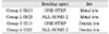
Prepared specimens were randomly divided into 4 groups. 4 experimental setups were used to simulate two different levels of cavity wall property (metal and dentin iris) and two different materials (ONE-STEP® and ALL-BOND® 2; Bisco Inc.) for each wall property. Metal iris with Teflon coating represented no bond to cavity wall, and dentin iris treated with bonding agent represented an optimal bond to cavity wall (Table 1).
Group M1 (ONE-STEP® & metal iris)
The surfaces were kept moist under blotting tissue. Two coats of One Step® were applied to the entire surface and thoroughly dry all surfaces for 10 seconds. ONE-STEP® was then light-cured for 10 s (Ultra Plus [Light intensity; 517 ± 55 mW/cm2]; Benlioglu Dental Inc., Binnaz Sk. 1 / 6 Kavaklidere, Ankara Turkey). A metal iris, which is 4 mm in diameter and 1.5 mm in height, was used as a mold for the composite. Teflon coating on the iris prevented it from adhering to the treated dentin. The iris, placed in a holder, was pressed against the treated dentin surface and the cavity was filled with a composite resin (RENEW®; Bisco Inc.), which was then irradiated for 40 s. The assembly was allowed to sit for an additional 4 min, and then immersed in distilled water at room temperature.
Group M2 (ALL-BOND® 2 & metal iris)
The surfaces were kept moist with the same blotting method as Group M1. Five coats of the mixed primer were applied to the dentin surface and lightly air dried for 5 s with oil free air. A thin layer of light cured unfilled bonding resin was applied to the primed surface. Primer and resin were then light-cured for 20 s. The procedure continued then as described for Group M1.
Group D1 (ONE-STEP® & dentin iris) and D2 (ALL-BOND® 2 & dentin iris)
The procedure as described for Group M was followed except for dentin iris treatment below. Dentin iris was made by sectioning 1.5 mm thick disk from the middle portion of the crown, and drilled a hole of 4 mm in diameter. Teflon tape, 0.14 mm thick, was attached to dentin iris. Before positioning the iris on the dentin surface, inner surface of the hole in dentin iris was etched and light-cured after applying bonding resin as described above.
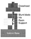
All specimens were stored in distilled water at room temperature (22℃) for 24 h before testing. They were then loaded in shear mode in an universal testing machine (model 4400; Instron Corp. Canton, MA, USA) at a crosshead speed of 0.5 mm/min by using a blunt blade covering the full edge of the iris. Techniques are shown schematically in Figure 1. After debonding, fracture analysis was performed using a 31.25 power microscope (OMPI pico(dental); Carl Zeiss, Jena, Germany) and the steel images from both sides of fracture surfaces were captured. Then the failure modes were classified as adhesive, cohesive in dentin, cohesive in the composite resin, or a combination thereof. The fractured surfaces of selected debonded specimens from each group were then examined further under scanning electron microscope (JSM-840A; JEOL Ltd, Tokyo, Japan). The shear bond strength was computed by dividing the maximum applied force by the bonded cross sectional area that was measured by Sigma Scan (Image ver 1.20; Jandel Scientific, Chicago, IL, USA) using the captured images. Data was evaluated statistically by a two-way ANOVA and t-test (M1 & D1, M2 & D2) using Sigma Stat (ver 2.03; Jandel Scientific).
III. Results
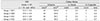
The shear bond strength and the failure modes are displayed in Table 2. The shear bond strength of the metal groups was significantly higher than those of the dentin iris groups (two-way ANOVA, p = 0.034). But, there was no significant difference between the bonding agent systems (two-way ANOVA, p = 0.263) and was no enough interaction between iris method and the bonding agent system (two-way ANOVA, p = 0.091). In ONE-STEP® groups, the shear bond strength of metal group was significant higher than those of dentin iris group (p = 0.005), but not in ALL-BOND® 2 (p = 0.774).
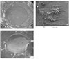
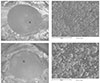
After verifying the failure modes of representative fractured surfaces using SEM (Figures 2 and 3), the failure modes of each specimen were classified under a microscope. The failure modes were mostly adhesive, and cohesive failures in dentin and in the composite resin were observed in a very rest-ricted area.
IV. Discussion
The area of the fractured surface varied between the two iris methods. Dentin iris method had more variation than metal iris method and this may be due to a lower modulus and more irregular shape of the dentin iris than the metal iris. Most areas of fractured surfaces observed in both methods were greater than the hole size. This might be due to the fact that the bonding agents were coated on the entire dentin area. Van Noort et al.13) had reported a two fold increase in shear bond strengths when coating the entire dentin area over those measured on a constrained area. Thus, in the iris method, the area must be measured after debonding and the shear bond strength should be calculated by dividing the maximum applied force by the measured area.
Under the specific condition of this study, our hypothesis was partly accepted, that is, the shear bond strength was affected by the cavity wall property represented by the two iris methods (two-way ANOVA; p = 0.034), especially in the case using ONE-STEP® (t-test; p = 0.005). However, between the groups using ALL-BOND® 2, our hypothesis was rejected due to insignificant difference (t-test; p = 0.774). The inconsistency in our results might come from the difference in the thickness of the adhesive layer being induced by the two bonding systems. Increasing the thickness of the adhesive layer was reported to relieve the shrinkage stress from the polymerizing composite resin14). This stress-relieving effect of thick adhesive layer might alleviate the difference in the influence of the wall property, representing C-factor, between the two iris methods using the two-bottle dentin adhesive system.
Finite element analysis has been used to describe the distribution of interfacial shear stresses5,8,9,13). From these it appears that the interfacial shear stresses are highly non-uniform and strongly influenced by bonding geometry, loading conditions and the mismatch in elasticity among the bonded layers.
In this study, iris method reduced the cohesive failure in the dentin and the composite resin. This is partly in agreement with data presented by Dickens et al11). They reported that iris method demonstrated fewer cohesive failures in dentin than conventional method, but no difference of cohesive failure in the composite resin. In this study, as the blade applying load onto the side of the iris was flat and the unbonded side of the iris was supported by a slide glass during loading, it might reduce the bending moment. As a result, the frequency and size of cohesive failures in the fractured surfaces of both the dentin and the composite resin were fewer and smaller than Dickens11). From the SEM images of the fractured surfaces, as the dentin surface and the corresponding surface of the composite resin were covered with glossy adhesive resin, it might assume that the fracture was happened within the adhesive layer. However, in the case of dentin iris, the rough surfaces, which had more open dentinal tubules, were observed more frequently (Figure 2). This finding was in a good agreement with the fracture patterns reported by Armstrong15) and Yoshikawa16). Therefore, it might be suggested that the shrinkage stress from the polymerizing composite resin under higher C-factor might cause the interfacial gap between the dentin and the adhesive layer and these defects might cause a decrease in shear bond strength during testing.
Many other studies have reported higher values of shear bond strength than this study. But in this study the absence of cohesive failures in dentin or the composite that have superior mechanical property might be suggested the reason for the reduction of the absolute values. So this study's result is thought to be close to the pure shear bond strength. In conclusion, pure adhesive failure is achieved by iris method. If the adhesive layers were thin, dentin iris method showed lower shear bond strength than metal iris method. So shear bond strength is thought to have relation with cavity wall property.
 XML Download