

 PDF
PDF ePub
ePub Citation
 Print
Print


Abstract
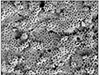
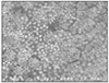
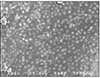
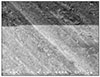
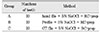
The purpose of this study was to evaluate the efficiency of the preparation of oval canals using hand and engine-driven instruments with SEM observation. Thirty single-rooted teeth with oval canal were used in this study. The teeth were divided into 3 groups. In group A, the teeth were instrumented up to a size 35 K-file using RC-prep and irrigated with 5% NaOCl between each file size. In group B, the teeth were instrumented with Profile according to the manufacture's instructions using RC-Prep and irrigated with 5% NaOCl between each file size. In group C, the teeth were instrumented with GT file according to the manufacture's instructions using RC-prep and irrigated with 5% NaOCl between each file size. Then, in all teeth, a final flush of 5ml of distilled water delivered for 30s. Canals were dried with sterile standardized paper points. After preparing the canals, the teeth were sectioned along their mesial and diatal surfaces by using low-speed diamond disc, chisel and mallet. Each root section was then dehydrated in graded concentration of alcohol (70, 80, 90, 100%), mounted on an aluminum stub, sputter-coated with gold-palladium and observed with scanning electron microscope (HITACHI S-4200) in middle and apical area.
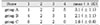
The results of this study were as follows:
In the middle area, group B and group C showed less smear layer than group A, and it was statistically significant (p < 0.05).
In the middle area, group B showed greater smear layer than group C, but it was not statistically significant (p > 0.05).
In the apical area, group C showed less smear layer than group A, and it was statistically significant (p < 0.05).
In the apical area, group A showed greater smear layer than group B, but it was not statistically significant (p > 0.05).
In the apical area, group B showed greater smear layer than group C, but it was not statistically significant (p > 0.05).
In all groups, the middle area was less smear layer than the apical area, and it was statistically significant (p < 0.05).
Figures and Tables

References
1. Esposito PT, Cunningham CJ. A comparison of canal preparation with nickel-titanium and stainless steel instruments. J Endod. 1995. 21:173–176.

2. Wu MK, Wasselink PR. A primary observation on the preparation and obturation of oval canals. Int Endod J. 2001. 34:137–141.
3. Weiger R, ElAyouti A, Löst C. Efficacy of hand and rotary instruments in shaping oval root canals. J Endod. 2002. 28:580–583.
4. Walton RE. Histologic evaluation of different methods of enlarging the pulp canal space. J Endod. 1976. 2:304–311.
5. Siqueira JF Jr, Araujo MC, Garcia PF, Fraga RC. Histological evaluation of the effectiveness of five instrumentation technique for cleanig the apical third of root canals. J Endod. 1997. 23:499–502.
6. Barbizam JV, Luis F. Effectiveness of manual and rotary instrumentation technique for cleaning flattened root canal. J Endod. 2002. 28:365–366.
7. Bechelli C, Colafranceschi M. Scanning electron microscope study the efficacy of root canal wall debridement of hand versus Lightspeed instrument. Int Endod J. 1999. 32:484–493.
8. Gutmann JL, Saunders WP, Nguyen L, Guo IY, Saunders EM. Ultrasonic root-end preparation Part 1 SEM analysis. Int Endod J. 1994. 27:318–324.
9. Cohen S, Burns PC. Pathway of the pulp. 7th ed. St. Louis: CV Mosby.
10. McComb D, Smith DC. A primary scanning electron microscope study of root canal after endodontic procedores. J Endod. 1975. 1:238–242.
11. Walton R, Torabinejad M. Principle and practice of endodontics. 1996. 2nd ed. Philadelphia: 212–214.
12. Brännström M, Nordenvall KJ, Glanz PO. The effect of EDTA-containing surface-active solutions on the morphology of prepared dentin: an vivo study. J Dent Res. 1980. 59:1127–1131.
13. Aktener BO, Cengiz T, Piskin B. The penetration of smear layer material into dentinal tubules during instrumentation with surface active reagents: a scanning electron microscope study. J Endod. 1989. 15:588–590.
14. Mader CL, Baumgartner JC, Peters DD. Scanning electron microscopic investigation of smeared layer on root canal walls. J Endod. 1984. 10:477–483.
15. Williams S, Goldman M. Penetrability of the smeared layer by strain of proteus vulgaris. J Endod. 1985. 11:385–388.
16. Dippel H, Borggreven J, Happenbrouvers P. Morphology and permeability of the dentin smear layer. J Prosthet Dent. 1984. 52:657–662.
17. Akpata ES, Blechman H. Bacterial invasion of pulpar dentin wall in vitro. J Dent Res. 1982. 61:435–438.
18. Nissan R, Sehal H, Pashley DH, Trowbridge H. Ability of bacterial endotoxin to permeate human dentin. J Endod. 1995. 21:62–64.
19. Brännström M, Nyborg H. Bacterial growth and pulpal changes under inlays cemented with zinc phosphate and epoxylate CBA 9080. J Prosthet Dent. 1974. 31:556–565.
20. Gençoğlu N, Samni S, Gunday M. Evalutation of sealing properties of Thermafil and Ultrafil techniques in the abscence or presence of smear layer. J Endod. 1993. 19:599–603.
21. Karagöz-Küçükay I, Bayirli G. An apical leakage study in the presence and abscence of smear layer. Int Endod J. 1994. 27:87–93.
22. Kennedy WA, Walker WA, Gough RW. Smear layer removal effect on apical leakage. J Endod. 1986. 12:21–27.
23. Cergneux M, Ciucchi B, Dietschi JM, Holz J. The influence of smear layer on the sealing ability of canal obturation. Int Endod J. 1987. 20:228–232.
24. Baumgartner JC, Mader CL. A scanning electron microscopic evaluation of four root canal irrigation regimens. J Endod. 1987. 13:147–157.
25. Grossman LI, Melman B. Solution of pulp tissue by chemical agents. J Am Dent Assoc. 1941. 28:223.
26. Weine FS. Endodontic therapy. 1996. 5th ed. St. Lous: Mosby.
27. Hülsmann M, Rummelin C, Schafers F. Root canal cleanliness after preparation with different endodontic handpiece and hand instruments: a comparative SEM investigation. J Endod. 1997. 23:301–306.
28. Heard F, Walton RE. Scanning electron microscope study comparing four root canals preparation techniques in small curved canals. Int Endod J. 1997. 30:323–331.
29. Bertrand MF, Pizzaardini P. The removal of the smear layer using the Quantec system. A study using the scanning electron microscope. Int Endod J. 1999. 32:217–224.
30. Alaçam T. Scanning electron microscope study comparing the efficacy of endodontic irrigating systems. Int Endod J. 1987. 20:287–294.
31. Ciucchi B, Khettabi M, Holz J. The effectiveness of different endodontic irrigation procedures on the removal of the smear layer: a scanning electron microscopic study. Int Endod J. 1989. 22:21–28.
32. Peters OA, Barbakow F. Effects of irrigation on debris and smear layer on canal walls prepared by two rotary techniques: A scanning electron microscopic study. J Endod. 2000. 26:6–10.
33. Gambarini G, Laszkiewicz J. A scanning electron microscopic study of debris and smear layer remaining following use of GT rotary instruments. Int Endod J. 2002. 35:422–427.
 XML Download