

 PDF
PDF ePub
ePub Citation
 Print
Print


Abstract
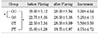
The purpose of this study was to compare the initial apical file(IAF) first file that fits to the apex in each canal before and after early flaring to analyze if the size of file to fit to the apex would increase after flaring. Eighty anterior teeth with complete apical formation and patent foramens were selected. The samples were randomly divided into 4 groups(GG, OS, GT, PT Group) of 20 teeth each. A file was fit to the apex in each canal and that size recorded. Radicular flaring were completed using different types of instruments. After flaring a file was again fit to the apex in the same manner as before and its size recorded.
The results of this study were as follows:
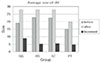
1. The mean diameter of IAF before flaring(file diameters in mm×10-2) was 19.81±8.32 before and 25.94±9.21 after(p<0.05).
2. The increase in diameter of IAF was approximately one file size for all groups.
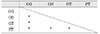
3. Ranking of increasing diameter of IAF were GG>GT>OS>PT group. There was a statistically significant difference between before and after flaring(p<0.05).
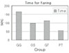
4. Ranking of the time for flaring were GG>GT>OS>PT group. There was a statistically significant difference between GG group and other groups(p<0.05).
5. In the case without change of IAF diameter, they showed decrease in force after flaring when IAF was pulled out from root canal(p<0.05).
This study suggested that early radicular flaring increases the file size that is snug at the apex, and awareness of that difference gives the clinician a better sense of canal size. Early flaring of the canal provides better apical size information and with this awareness, a better decision can be made concerning the appropriate final diameter needed for complete apical shaping.
Figures and Tables


References
1. Cohen S, Burns R. Pathways of the pulp. 1994. 6th ed. St. Louis: CV Mosby;179–218.
2. Lim SS. Clinical Endodontics. 1999. 2nd ed. Seoul: Uichihaksa;128–136.
3. Grossman L. Endodontic practice. 1985. 10th ed. Philadelphia: Lea & Febiger;207.
4. Weine FS. Endodontic therapy. 1982. 3rd ed. St. Louis: Mosby Co;256–340.
5. Ingle J, Bakland L. Endodontics. 1994. 4th ed. Philadelphia: Lea & Febiger;92–227.
6. Oliet S, Sorin SM. Cutting efficiency of endodontic Remers. Oral Surg Oral Med Oral Pathol. 1973. 36:243.
7. Walton R, Torabinejad M. Principles and practice of endodontics. 1989. 1st ed. Philadelphia, USA: WB Saunders Co;189–190.
8. Leeb J. Canal orifice enlargement as related to biomechanical preparation. J Endod. 1983. 9:463–470.

9. Goerig AC, Michelich RJ, Schultz HH. Instrumentation of root canals in molar using the step-down technique. J Endod. 1982. 8:550–554.
10. Morgan LF, Montgomery S. An evaluation of the crown-down pressureless technique. J Endod. 1984. 10:491–498.
11. Fava LR. The double-flared technique: an alternative for biomechanical preparation. J Endod. 1983. 9:76–80.
12. Abou-Rass M, Jastrab RJ. The use of rotary instruments as auxiliary aids to root canal preparation of molars. J Endod. 1982. 8:78–82.
13. Isom TL, Marshall JG, Baumgartner JC. Evaluation of root thickness in curved canals after flaring. J Endod. 1995. 21:368–371.
14. Contreras MA, Zinman EH, Kaplan SK. Comparison of the First File that Fits at the Apex, Before and After Early Flaring. J Endod. 2001. 27:113–116.
15. Musikant BL, Cohen BI, Deutsch AS. The evolution of instrumentation and obturation leading to as simplified approach. Compend Contin Educ Dent. 2000. 21:980–990.
16. Kessler JR, Peters DD, Lorton S. Comparision of the relative risk of molar root perforations using various endodontic instrumentation techniques. J Endod. 1983. 9:439–447.
17. al-Omari MA, Dummer PMH. Canal blockage and debris extrusion with eight preparation techniques. J Endod. 1995. 21:154–158.
18. Coldero LG, McHugh S, MacKenzie D, Saunders WP. Reduction in intracanal bacteria during root canal preparation with and without apical enlargement. Int Endod J. 2002. 35:437–446.
19. Fogarty TJ, Montgomery S. Effect of preflaring on canal transportation. Oral Surg Oral Med Oral Pathol. 1991. 72:345–350.
20. Lambrianidis T, Tosounidou E, Tzoanopoulou M. The effect of maintaining apical patency on periapical Extrusion. J Endod. 2001. 27:696–698.
21. Swindle RB, Neaverth EJ, Pantera EA, Ringle RD. Effect of coronal-radicular flaring on apical Transportation. J Endod. 1991. 17:147–149.
22. Rollison S, Barnett F, Stevens RH. Efficacy of bacterial removal from instrumented root canals in vitro related to instrumentation technique and size. Oral Surg Oral Med Oral Pathol Oral Radiol Endod. 2002. 94:366–371.
23. Davis RD, Marshall JG, Baumgartner JC. Effect of early coronal flaring on working length change in curved canals using rotary nickel-titanium versus stainless steel instruments. J Endod. 2002. 28:438–442.
24. Gluskin AH, Brown DC, Buchanan LS. A reconstructed computerized tomographic comparison of Ni-Ti rotary GT files versus traditional instruments in canals shaped by novice operators. Int Endod J. 2001. 34:476–484.
25. Tan BT, Messer HH. The quality of apical canal preparation using hand and rotary instruments with specific criteria for enlargement based on initial apical file size. J Endod. 2002. 28:658–664.
26. Roane JB, Sabala CL, Duncanson MG. The "Balanced Force" concept for instrumentation of curved Canals. J Endod. 1985. 11:203–211.
27. Goldberg F, Massone EJ. Patency file and apical transportation: An in vitro study. J Endod. 2002. 28:510–511.
28. Gani O, Visvisian C. Apical canal diameter in the first upper molar at various ages. J Endod. 1999. 25:689–691.
29. Fuge KN, Stuck AMC, Love RM. A comparison of digitally scanned radiographs with conventional film for the detection of small endodontic instruments. Int Endod J. 1998. 31:123–126.
 XML Download