

 PDF
PDF ePub
ePub Citation
 Print
Print


Abstract
Complete understanding of the exterior and interior structure of the tooth would be prerequisite to the successful clinical results, especially in the restorative and endodontic treatment.
Although three-dimensional reconstruction method using x-ray microtomography could not be used in clinical cases, it may be the best way to reconstruct the morphologic characteristics of the tooth structure in detail without destructing the tooth itself. This study was done to three dimensionally reconstruct every teeth in the arch in order to increase the understanding about the endodontic treatment and to promote the effective restorative treatment by upgrading the knowledge of the tooth morphology.
After placing tooth between the microfocus x-ray tube and the image intensifier to obtain two-dimensional images of each level, scanning was done under the condition of 80 keV, 100 µA, 16.8 magnification with the spot size of 8 µm. Cross-section pixel size of 16.28 µm and 48.83 cross-section to cross-section distance were also used.
From the results of this study, precise three dimensional reconstructed images of every teeth could be obtained. Furthermore, it was possible to see image that showed interested area only, for example, enamel portion only, pulp and dentin area without enamel structure, pulp only, combination image of enamel and pulp, etc.
It was also possible to see transparent image without some part of tooth structure. This image might be used as a guide when restoring and preparing the full and partial crown by showing the positional and morphological relationship between the pulp and the outer tooth structure.
Another profit may be related with the fact that it would promote the understanding of the interior structure by making observation of the auto-rotating image of .AVI file from the various direction possible.
Figures and Tables
Fig. 1
Three-dimensionally reconstructed upper anteriors. Upper: central incisor, Middle: lateral incisor, Lower: canine. From left to right: labial, lingual, mesial, distal, incisal, apical view
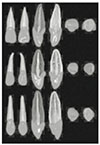
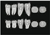
Fig. 2
Three-dimensionally reconstructed upper premolars. Upper: 1st premolar, Lower: 2nd premolar. From left to right: labial, lingual, mesial, distal, incisal, apical view
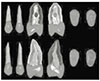
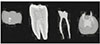
Fig. 3
Three-dimensionally reconstructed upper molars. Upper: 1st molar, Lower: 2nd molar. From left to right: labial, lingual, mesial, distal, incisal, apical view
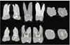
Fig. 4
Three-dimensionally reconstructed lower anteriors. Upper: central incisor, Middle: lateral incisor, Lower: canine. From left to right: labial, lingual, mesial, distal, incisal, apical view
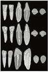
Fig. 5
Three-dimensionally reconstructed lower premolars. Upper: 1st premolar, Lower: 2nd premolar. From left to right: labial, lingual, mesial, distal, incisal, apical view
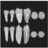
Fig. 6
Three-dimensionally reconstructed lower molars. Upper: 1st molar, Lower: 2nd molar. From left to right: labial, lingual, mesial, distal, incisal, apical view
References
1. Bellizzi R, Hartwell G. Radiographic evaluation of root canal anatomy of in vivo endodontically treated maxillary premolars. J Endod. 1985. 11:37–39.

2. Kulild JC, Peters DD. Incidence and configuration of canal systems in the mesiobuccal root of maxillary first and second molars. J Endod. 1990. 16:311–317.
3. Tagger M. Clearing of teeth for study and demonstration of the pulp. J Dent Educ. 1976. 40:172–174.
4. Melton DC, Krell KV, Fuller MW. Anatomical and histological features of C-shaped canals in mandibular second molars. J Endod. 1991. 17:384–388.
5. Hirano Y, Aoba T. Computer-assisted reconstruction of enamel fissures and carious lesions of human molars. J Dent Res. 1995. 74:1200–1205.
6. Jung EH, Shin DH. Morphologic analysis of C-shaped root using 3D reconstruction. J Korean Acad Conserv Dent. 2002. 27(4):421–431.
7. Elliott JC, Dover SD. Three-dimensional distribution of mineral in bone at a resolution of 15 micron determined by x-ray microtomography. Metab Bone Dis Relat Res. 1984. 5:219–221.
8. Nielsen RB, Alyassin AM, Peters DD, Carnes DL, Lancaster J. Microcomputed tomography: an advanced system for detailed endodontic research. J Endod. 1995. 21:561–568.
9. Dowker SEP, Davis GR, Elliott JC. X-ray microtomography: Nondestructive three-dimensional imaging for in vitro endodontic studies. Oral Surg Oral Med Oral Pathol Oral Radiol Endod. 1997. 83(4):510–516.
10. Bjorndal L, Carlsen O, Thuesen G, Darvann T, Kreiborg S. External and internal macromorphology in 3D-reconstructed maxillary molars using computerized x-ray microtomography. Int Endod J. 1999. 32:3–9.
11. Lyroudia K, Nikolaidis N, Pitas I, Palakidis K. Three computer methods to reconstruct pulpal blood vessels and nerves. J Endod. 1995. 21:501–504.
12. Sennerby L, Wennerberg A, Pasop F. A new microtomographic technique for non-invasive evaluation of the bond structure around implants. Clin Oral Implants Res. 2001. 12:91–94.
13. Tachibana H, Matsumoto K. Applicability of X-ray computerized tomography in endodontics. Endod Dent Traumatol. 1990. 6:16–20.
14. Bergmans L, Cleynenbreugel JV, Wevers M, Lambrechts P. A methodology for quantitative evaluation of root canal instrumentation using microcomputed tomography. Int Endod J. 2001. 34:390–398.
 XML Download