

 PDF
PDF ePub
ePub Citation
 Print
Print


Abstract
There are increasing usage of Nickel-Titanium rotary files in modern clinical endodontic treatment because it is effective and faster than hand filing due to reduced step.
This study was conducted to evaluate the effect of canal preparations using 3 different rotary Nickel-Titanium files that has different cross sectional shape and taper on the maintenance of canal curvature. Simulated resin block were instrumented with Profile(Dentsply, USA), GT rotary files(Dentsply, USA), Hero 642(Micro-Mega, France), and Pro-Taper(Dentsply, USA).
The image of Pre-instrumentation and Post-instrumentation were acquired using digital camera and overspreaded in the computer. Then the total differences of canal diameter, deviation at the outer portion of curvature, deviation at the inner portion of curvature, movement of center of the canal and the centering ratio at the pre-determined level from the apex were measured.
Results were statistically analyzed by means of ANOVA, followed by Scheffe test at a significance level of 0.05.
The results were as follows;
1. Deviation at the outer portion of curvature, deviation at the inner portion of curvature were showed largest in Pro-Taper, so also did in the total differences of canal diameter(p<0.05).
2. All the groups showed movements of center. Profile combined with GT rotary files and Hero 642 has no difference but Pro-Taper showed the most deviation(p<0.05).
3. At the 1, 2, 3mm level from the apex movements of center directed toward the outer portion of curvature, but in 4, 5 mm level directed toward the inner portion of curvature(p<0.05).
As a results of this study, it could be concluded that combined use of other Nickel-Titanium rotary files is strongly recommended when use Pro-Taper file because it could be remove too much canal structure and also made more deviation of canal curvature than others.
Figures and Tables
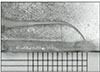
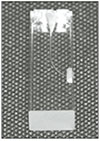 | Fig. 1A transluscent Endo-training block used in study that has single 35° - curvature measured by Schneider method.
|
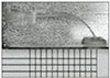
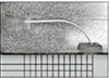
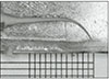
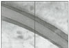 | Fig. 6The extent of the change toward outer portion of canal curvature is the area from the line before canal shaping to the outer line, and the area below the line is the extent of the change toward inner part.
|


Table 3
Comparison among test groups of changes in total cross sectional length of canal of each group at measurement site

![]()

Table 5
Comparison among test groups of deviations at the outer portion of curvature of each group at measurement site

![]()

Table 7
Comparison among test groups of deviations at the inner portion of curvature of each group at measurement site

![]()


References
1. Schilder H. Cleaning and shaping the root canal. Dent Clin North Am. 1974. 18(2):269–296.
2. Wildey WL, Senia ES, Montgomery S. Another look at root canal instrumentation. Oral Surg Oral Med Oral Pathol. 1992. 74(4):499–507.

3. Skidmore AE, Bjorndal AM. Root canal morphology of the human mandibular first molar. Oral Surg Oral Med Oral Pathol. 1971. 32(5):778–784.
4. Abou-Rass M, Frank AL, Glick DH. The anticurvature filing method to prepare the curved root canal. J Am Dent Assoc. 1980. 101(5):792–794.
5. Civjan S, Huget EF, DeSimon LB. Potential applications of certain nickel-titanium (nitinol) alloys. J Dent Res. 1975. 54(1):89–96.
6. Stoeckel D, Yu W. Superelastic Ni-Ti wire. Wire J Int. 1991. 3:45–50.
7. Glossen CR, Haller RH, Dove SB, del Rio CE. A comparison of root canal preparations using Ni-Ti hand, Ni-Ti engine-driven, and K-Flex endodontic instruments. J Endod. 1995. 21(3):146–151.
8. Serene TP, Adam JD, Saxena A. Nickel-titanium instruments : Applications in Endodontics. St Louis, Mosby, USA: Ishiyaku Euro America, Inc.;1–110.
9. Abou-Rass M, Frank AL, Glick DH. The anticurvature filing method to prepare the curved root canal. J Am Dent Assoc. 1980. 101(5):792–794.
10. Thompson SA, Dummer PM. Shaping ability of Hero 642 rotary nickel-titanium instruments in simulated root canals: Part 2. Int Endod J. 2000. 33(3):255–261.
11. Schafer E. Shaping ability of Hero 642 rotary nickel-titanium instruments and stainless steel hand K-Flexofiles in simulated curved root canals. Oral Surg Oral Med Oral Pathol Oral Radiol Endod. 2001. 92(2):215–220.
12. Thompson SA, Dummer PM. Shaping ability of Mity Roto 360 degrees and Naviflex rotary nickel-titanium instruments in simulated root canals. Part 1. J Endod. 1998. 24(2):128–134.
13. Luiten DJ, Morgan LA, Baugartner JC, Marshall JG. A comparison of four instrumentation techniques on apical canal transportation. J Endod. 1995. 21(1):26–32.
14. Gambill JM, Alder M, del Rio CE. Comparison of nickel-titanium and stainless steel hand-file instrumentation using computed tomography. J Endod. 1996. 22(7):369–375.
15. Weine FS. "Endodontic Therapy". 1996. 5th ed. St. Louis: Mosby, Inc.;330–331.
16. Lim KC, Webber J. The validity of simulated root canals for the investigation of the prepared root canal shape. Int Endod J. 1985. 18:240–246.
17. Thompson SA, Dummer PM. Shaping ability of Hero 642 rotary nickel-titanium instruments in simulated root canals: Part 1. Int Endod J. 2000. 05. 33(3):248–254.
18. Elliott LM, Curtis RV, Pitt Ford TR. Cutting pattern of nickel - titanium files using two preparation techniques. Endod Dent Traumatol. 1998. 14(1):10–15.
19. Bryant ST, Thompson SA, al-Omari MA, Dummer PM. Shaping ability of ProFile rotary nickel-titanium instruments with ISO sized tips in simulated root canals: Part 2. Int Endod J. 1998. 31(4):282–289.
20. Roane JB, Sabala CL, Duncanson MG Jr. The "balanced force" concept for instrumentation of curved canals. J Endod. 1985. 11(5):203–211.
 XML Download