

 PDF
PDF ePub
ePub Citation
 Print
Print


Abstract
The objectiveness of this study was to evaluate whether low-viscosity composite can bond effectively to dentin surface without bonding resin. The low-viscosity composites being 50wt% filler content were made by the inclusion of bonding resin of two self-etching systems(Clearfil SE Bond, Unifil Bond) varied with contents as 0, 10, 20, 30, 40, 50wt%.
Exposed dentin surfaces of extracted 3rd molars are used. Dentin bond strengths were measured. The tests were carried out with a micro-shear device placed testing machine at a CHS of 1mm/min after a low-viscosity composite was filled into an iris cut from micro tygon tubing with internal diameter approximately 0.8mm and height of 1.0mm.
Flexural strength and modulus was increased with the addition of bonding resin.
Micro-shear bond strength to dentin was improved according to content of bonding resin irrespective of applying or not bonding resin in bonding procedure, and that of Clearfil SE Bond groups was higher than Unifil Bond.
There were no significant difference whether use of each bonding resin in bonding procedure for S-40, S-50, U-50(p>0.05).
In SEM examination, resin was well infiltrated into dentin after primed with self-etching primer only for S-50 and U-50 in spite of the formation of thinner hybrid layer.
Low viscosity composite including some functional monomer may be used as dentin bonding resin without an intermediary bonding agent. It makes a simplified bonding procedure and foresees the possibility of self-adhesive restorative material.
Figures and Tables
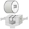
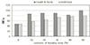
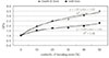
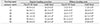
Fig. 4
Comparison between bond strength with or without applying bonding resin for Clearfil SE Bond.
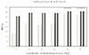
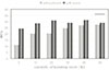
Fig. 6
SEM image of interfaces bonded with Clearfil SE Bond. (a) as the manufacturer's instruction and bonded with S-0 resin. Note the well-formed resin tags with lateral branches and homogeneous thicker hybrid layer. (b) Specimen primed with SE Primer without bonding resin applied and bonded with S-0 resin directly. An inconsistent and thinner hybrid layer and a decreased number of tags were formed.
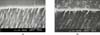
Fig. 7
The adhesive interfaces with Clearfil SE Bond. (a) as the manufacturer's instruction and bonded with S-20 resin. Hybrid layer was formed uniformly and resin tags were infiltrated into dentinal tubules deeply. (b) Specimen primed with SE Primer without bonding resin applied and bonded with S-20 resin directly. Note few tags relatively and uneven hybrid layer.
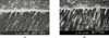
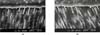
Fig. 8
The adhesive interfaces with Clearfil SE Bond. (a) as the manufacturer's instruction and bonded with S-50 resin. Hybrid layer was well-formed uniformly and a number of resin tags and canaliculi were observed. (b) Specimen primed with SE Primer without bonding resin applied and bonded with S-50 resin directly. In spite of thinner hybrid layer relatively, the presence of a great number of long tags were observed.
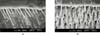
Fig. 9
SEM image of interfaces bonded with Unifil Bond. (a) as the manufacturer's instruction and bonded with U-0 resin. Note uniform hybrid layer but the poorly-formed resin tags. (b) Specimen primed with self-etching primer of Unifil Bond without bonding resin applied and bonded with U-0 resin directly. Hybrid layer was thin and inconsistent and there was scarcely any tag.
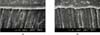
Fig. 10
SEM image of interfaces primed with self-etching primer of Unifil Bond and bonded with U-20 resin replacing the bonding resin. (a) Note resin infiltrated into dentinal tubule very well and lateral braches at 1,500× of magnification. (b) Magnified(×6,000) image of white box area in (a) micrograph. The hybrid layer is approximately 1~2µm thick, the resin tags exhibit some few tags relatively thinner hybrid layer.
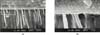
Fig. 11
The adhesive interface with Unifil Bond. (a) as the manufacturer's instruction and bonded with U-50 resin. Hybrid layer was well-formed uniformly and a number of resin tags were observed. (b) Specimen primed with self-etching primer of Unifil Bond without bonding resin applied and bonded with U-50 resin directly. In spite of thinner hybrid layer relatively, the presence of a great number of long tags were observed.


References
1. Ferracane JL. Current trends in dental composites. Crit Rev Oral Biol Med. 1995. 6:302–318.

2. Perdigao J, Lambrechts P, van Meerbeek B, Bream M, Yildiz E, Yucel T, Vanherle G. The interaction of adhesive systems with human dentin. Am J Dent. 1996. 9:167–173.
3. Davidson CL, De Gee AJ, Feilzer AJ. The competition between the composite-dentin bond strength and the polymerization contraction. J Dent Res. 1984. 63(12):1396–1399.
4. Buonocore MG. A simple method of increasing the adhesion of acrylic filling materials to enamel surfaces. J Dent Res. 1955. 34:849–854.
5. Perdigão J, Frankenberger R, Rosa BT, Breschi L. New trends in dentin/enamel adhesion. Am J Dent. 2000. 13:25D–30D.
6. Hulmes DJ, Wess TJ, Prockop DJ, Fratzl P. Radial packing, order and disorder in collagen fibrils. Biophys J. 1995. 68:1661–1670.
7. Nakabayashi N, Pashley DH. Hybridization of dental hard tissues. 1998. Quintessence Publishing Co;1–107.
8. van Meerbeek B, Perdigao J, Lambrechts P, Vanherle G. The clinical performance of adhesives. J Dent. 1998. 26:1–20.
9. Fritz UB, Finger WJ, Stean H. Salivary contamination during bonding procedures with a one-bottle adhesive system. Quintessence Int. 1998. 29(9):567–572.
10. Hayakawa T, Kikutake K, Nemoto K. Conformational and quantum analysis of dental adhesive carboxylic acid and carboxylic acid anhydride monomers. Dent Mater J. 2001. 20(1):1–15.
11. Tay FR, Gwinnett AJ, Pang KM, Wei SH. Resin permeation into acid-conditioned, moist and dry dentin: a paradigm using water-free adhesive primers. J Dent Res. 1996. 75(4):1034–1044.
12. Prati C, Chersoni S, Mongiorgi R, Pashley DH. Resin-infiltrated dentin layer formation of new bonding systems. Oper Dent. 1998. 23:185–194.
13. Choi KK, Kim SW, Choi HY. Effect of filler addition to bonding agents on the physical properties and the bond strength to bovine teeth. 2002. The First International Congress on Adhesive Dentistry(IAD, Program and Abstracts);434.
14. Miyazaki M, Platt JA, Onose H, Moore BK. Influence of dentin primer application methods on dentin bond strength. Oper Dent. 1996. 21:167–172.
15. Imai T, Itoh K, Tani C, Manabe A, Yamashita T, Hisamitsu H, Wakumoto S. Effectiveness of simplified dentin bonding systems. Dent Mater J. 1998. 17(1):1–10.
16. Bayne SC, Thompson JY, Swift EJ Jr, Stamatiades P, Wilkerson M. A characterization of first-generation flowable composites. J Am Dent Assoc. 1998. 129(5):567–577.
17. Feilzer AJ, De Gee AJ, Davidson CL. Quantitative determination of stress reduction by flow in composite restorations. Dent Mater. 1990. 6:167–171.
18. Unterbrink GL, Liebenberg WH. Flowable resin composites as "filled adhesives": Literature review and clinical recommendations. Quintessence Int. 1999. 30:249–257.
19. Choi SJ, Kim MJ, Kwon HC. Microleakage of posterior packable composite resin at the gingival margins of class II cavities. J Korean Acad Conserv Dent. 2002. 27(3):249–256.
20. Frankenberger R, Lopes M, Perdigao J, Ambrose WW, Rosa BT. The use of flowable composites as filled adhesives. Dent Mater. 2002. 18:227–238.
21. ISO 4049. International Standard Dentistry-Resin-based dental filling materials. 1988.
22. Kemp-Scholte CM, Davidson CL. Complete marginal seal of class V resin composite restorations effected by increased flexibility. J Dent Res. 1990. 69:1240–1243.
23. Choi KK, Condon JR, Ferracane JL. The effects of adhesive thickness on polymerization contraction stress of composites. J Dent Res. 2000. 79:812–817.
24. Montes MA, de Goes MF, da Cunha MR, Soares AB. A morphological and tensile bond strength evaluation of an unfilled adhesive with low-viscosity composites and a filled adhesive in one and two coats. J Dent. 2001. 29(6):435–441.
25. Tay FR, Moulding KM, Pashley DH. Distribution of nanofillers from a simplified-step adhesive in acid-conditioned dentin. J Adhes Dent. 1999. 1:103–117.
26. Perdigao J, Lambrechts P, van Meerbeek B, Tome AR, Vanherle G, Lopes AB. Morphological field emission-SEM study of the effect of six phosphoric acid etching agents on human dentin. Dent Mater. 1996. 12(4):262–271.
27. Willems G, Lambrechts P, Braem M, Celis JP, Vanherle G. A classification of dental composites according to their morphological and mechanical characteristics. Dent Mater. 1992. 8:310–319.
28. Braem M, Ringer W, van Doren V, Lambrechts P, Vanherle G. Mechanical properties and filler fraction of dental composites. Dent Mater. 1989. 5:346–349.
29. Lee YS, Choi KK, Park SJ. Effect of resin matrix on degree of conversion and fracture toughness of dental composites. J Korean Acad Conserv Dent. 2002. 27(1):77–86.
30. Shimada Y, Antonucci JM, Schumacher GE, McDonough WG, Tagami J. International symposium; Advanced adhesive dentistry; Effects of regional tooth structure and sectioning orientation on microshear bond strength. 1999. Kuraray Co. Ltd;91–103.
31. Sano H, Shono T, Sonoda H, Takatsu T, Ciucchi B, Carvalho RM, Pashley DH. Relationship between surface area for adhesion and tensile bond strength-evaluation of a micro-tensile bond tests. Dent Mater. 1994. 10:236–240.
32. Pashley DH, Carvalho RM, Sano H, Nakajima M, Yoshiyama M, Shono Y, Fernandes CA, Tay F. The microtensile bond test: a review. J Adhes Dent. 1999. 1(4):299–309.
33. Hasegawa T, Itoh K, Koike T, Yukitani W, Hisamitsu H, Wakumoto S, Fujishima A. Effect of mechanical properties of resin composites on the efficacy of the dentin bonding system. Oper Dent. 1999. 24(6):323–330.
34. Frankenberger R, Perdigao J, Rosa BT, Lopes M. "No-bottle" vs "multi-bottle" dentin adhesives--a microtensile bond strength and morphological study. Dent Mater. 2001. 17(5):373–380.
35. Nakabayashi N, Takarada K. Effect of HEMA on bonding to dentin. Dent Mater. 1992. 8:125–132.
36. Wang T, Nikaido T, Nakabayashi N. Photocure bonding agent containing phosphoric methacrylate. Dent Mater. 1991. 7(1):59–62.
37. Igarashi K, Toida T, Nakabayashi N. Effect of phenyl-P/HEMA primer on bonding to demineralization dentin by phosphoric acid. Dent Mater J. 1997. 16:55–60.
38. Hume R, Bayne SC, Duke ES. What is the future of amalgam? Quintessence Int. 1996. 27:136–141.
 XML Download