

 PDF
PDF ePub
ePub Citation
 Print
Print


Abstract
The purpose of this study was to evaluate the color changes of composite resin polymerized with three type of light curing units. Composite resin (Z100, shade A2) were applied in a cylindrical metal mold(2 mm thick, 7 mm diameter).
Twenty specimens according to light curing units were made.
Group1: the specimens were polymerized with Apollo 95E for 3seconds(1370 mW/cm2).
Group2: the specimens were polymerized with XL 3000 for 40seconds(480 mW/cm2).
Group3: the specimens were polymerized with Spectrum 800 for 10 seconds(250 mW/cm2) and 30 seconds(700 mW/cm2).
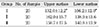
The microhardness values(VHN) of upper and lower surfaces specimens after light polymerization were measured for the degree of polymerization. All specimens were stored in distilled water at 60℃ for 30 days.
The color characteristics(L*, a*, b*) of the specimens before and after immersion were measured by spectrophotometer and the total color difference (ΔE*) was computed.
The results obtained were as follows:
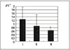
1. The microhardness values of Group I showed significantly lower than those of Group II and III(p<0.05).
2. In all groups the ΔE* values presented below 2.0.
3. Group I showed the highest ΔE* values followed order from highest to lowest by Group II and III (p<0.05).
Figures and Tables
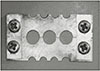
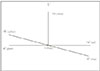
Fig. 3
Graphic representating of mean microhardness values(VHN) of each group
I: Apollo 95E, II: XL 3000, III: Spectrum 800
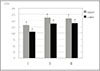
Fig. 4
Graphic representation of the chromatic color changes of group I, II and III produced by storing for 30 days in distilled water at 60℃
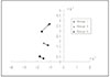
Fig. 5
Total color difference(ΔE*) in group I, II and III
I: Apollo 95E, II: XL 3000, III: Spectrum 800

References
1. Ruyter IE. Conversion in different depths of ultraviolet and visible light activated composite materials. Acta Odontol Scand. 1982. 40:179–182.

2. Shintani H, Inoue T, Yamaki M. Analysis of camphoroquinone invisible light cured composite resins. Dent Mater. 1985. 1:124–126.
3. Leung R, Fan P, Johnson W. Post-irradiation polymerization of visible light activated composite resin. J Dent Res. 1983. 62:363–365.
4. Friedman J. Care and maintenance of dental curing light. Dent Today. 1991. 10:40–41.
5. Carvalho RM, Pereira JC, Yoshiyama M, Pashley DH. A review of polymerization contraction: the incidence of stress development versus stress relief. Oper Dent. 1996. 21(1):17–24.
6. Uno S, Asmussen E. Marginal adaptation of restorative resin polymerized at a reduced rate. Scand J Dent Res. 1991. 99:440–444.
7. Unterbrink GL, Muessner R. Influence of light intensity on two restorative systems. J Dent. 1995. 23(3):183–189.
8. Mehl A, Hickel R, Kunzelmann KH. Physical properties and gap formation of light-cured composites with and without soft start-polymerization. J Dent. 1997. 25(3):321–330.
9. Koran P, Kurschner R. Effect of sequential versus continuous irradiation of a light-cured resin composite on shrinkage, viscosity, adhesion and degree of polymerization. Am J Dent. 1998. 11(1):17–22.
10. Peutzfeldt A, Sahafi A, Asmussen E. Characterization of resin composites polymerized with plasma arc curing units. Dent Mater. 2000. 16(5):330–336.
11. Hanyang University Department of Physics. Plasma Application. 2000. Laboratory.
12. Eldiwany M, Komatsu S, Powers JM. Curing light intensityaffects mechanicalproperties of composites. J Dent Res. 1997. 76:73.
13. Caughman WF, Caughman GB, Shiflett RA, et al. Correlation of cytotoxicity, filler loading and curing time of dental composites. Biomaterials. 1991. 12:737–740.
14. Seghi RR, Gritz MD, Kim J. Colorimetric changes in composites resulting fromvisible-light-initiated polymerization. Dent Mater. 1990. 6:133–137.
15. Brauer GM. Color changes of composites on exposure to various energy sources. Dent Mater. 1988. 4:55–59.
16. Powers JM, Barakat MM, Ogura H. Color and optical properties of posteriorcomposites resin restoration. Dent Mater J. 1985. 4:62–67.
17. Noie F, O'Keeefe KL, Powers JM. Color stability of resin cements after accelerated aging. Int J Prosthodont. 1995. 8:51–55.
18. Swift EJ, Hammel SA, Lund PS. Colorimetric evaluation of vita shade resin composites. Int J Prosthodont. 1994. 7:356–361.
19. Asmussen E. An accelerated test for color stability of restorative resins. Acta Odontol Scand. 1981. 39:329–332.
20. Roh BD, Park SH, Lee CS. An experimental study of the degree of conversion and cytotoxicity of dual cure resin cements. J Korean Acad Conserv Dent. 1995. 20(1):33–33.
21. DeWald JP, Ferracane JL. A comparison of four modes of evaluation depth of cure of light-activated composites. J Dent Res. 1982. 66:727–730.
22. Vargas MA, Cobb DS, Schmit JL. Polymerization of composite resins:Argon laser vs conventional light. Oper Dent. 1998. 23:87–93.
23. Sakaguchi RL, Sasik CT, Bunczak MA, Douglas WH. Strain gauge method for mearsuring polymerization contraction of composite restoratives. J Dent. 1991. 19(5):312–326.
24. Goracci G, et al. Curing light intensity and marginal leakage of resin composite restorations. Quintessence Int. 1996. 27(5):355–362.
25. Martin FE. A survey of the efficiency of visible light curing units. J Dent. 1998. 26(3):239–243.
26. Rueggeberg FA, Craig RC. Correlation of parameters used to estimate monomer conversion in a lightcured composite. J Dent Res. 1988. 67:932–937.
27. Asmussen E. Restorative resins:hardness and strengthvsquality of remaining double bonds. Scand J Dent Res. 1982. 90:484–489.
28. Hansen EK. After-polymerization of visible light activated resins ; surface hardness vs light source. Scand J Dent Res. 1983. 91:406–410.
29. Backer J, Dermaut L, Bruynooghe W. The depth of polymerization of visible light-cured composite resins. Quintessence Int. 1985. 10:693–699.
30. Leung RL, Kahn RL, Fan PL. Comparison of depth of polymerization evaluation method for photoactivated composite. J Dent Res. 1982. 61:300. IADR Abstract # 1095.
31. Munksgaard EC, Peutzfeldt A, Asmussen E. Elution of TEGDMA and BisGMA from a resin and a resin composite cured with halogen or plasma light. Eur J Oral Sci. 2000. 108(4):341–345.
32. Silikas N, Eliades G, Watts DC. Light intensity effects on resin-composite degree of conversion and shrinkage strain. Dent Mater. 2000. 16(4):292–296.
33. Asmussen E. Factor affecting the color stability of restorative resins. Acta Odontol Scand. 1983. 41:11–18.
34. Um CM, Ruyter IE. Staining of resin-based veneering materials with coffee and tea. Quintessence Int. 1991. 22:377–386.
35. Ruyter IE, Svendsen SA. Remaining methacrylate groups in composite restorative materials. Acta Odontol Scand. 1978. 36:75–82.
36. Hayshi H, Maejima K, Kezuka K, Ogushi K, Kono A, Fusayama TT. In vitro study of discoloration of composite resins. J Prosthet Dent. 1974. 32:66–69.
37. Dodge WW, Dale RA, Colley RL, Duke ES. Comparison of wet and dry finishing of resin composites with aluminium oxide discs. Dent Mater. 1991. 7(1):18–20.
38. Han WS, Kum KY, Lee CY. The influence of fluoride on remineralization of artificial dental caries. J Korean Dent Assoc. 1977. 15:1009–1012.
39. Gross MD, Moser JB. A colorimetric study ofcoffeeandtea staining of four composite resins. J Oral Rehabil. 1977. 4:311–322.
40. Satou N, Khan AM, Mastsumae I, Satou J, Shintani H. In vitro color change of composite-based resins. Dent Mater. 1989. 5:384–389.
41. Wozniak WT, Muller TP, Silverman R, Moser JB. Photographic assessment of colour changes in cold and heat-cure resins. J Oral Rehabil. 1981. 8:333–339.
42. Raptis CN, Powers JM, Fan PL, Yu R. Staining of composite resins by cigarette smoke. J Oral Rehabil. 1982. 9:367–371.
43. Ruyter IE, Nilner K, Moller B. Color stability of dental composite resin materials for crown and bridge veneers. Dent Mater. 1987. 13:246–251.
44. Seghi RR, Jonston WM, O'Brien WJ. Performance assessment of colorimetric devices on dental porcelain. J Dent Res. 1989. 68:1755–1759.
45. Dijken JWV. A clinical evaluation of anterior conventional microfilled and hybrid composite resin filling. Acta Odontol Scand. 1986. 44:357.
46. Kim CW, Lim BS, Moon HJ. Effect of Organic Solutions on the Surface Roughness and Color Stability of Dental Composite Resins. J Korea Res Soc Dent Mater. 1999. 26(1):17–34.
47. Dietchi D, Campanile G, Holz J, Meyer JM. Comparison of the color stability of ten new-generation composites : An in vitro study. Dent Mater. 1994. 10:353–362.
 XML Download