

 PDF
PDF ePub
ePub Citation
 Print
Print


Abstract
The purpose of this study was to compare the histomorphological change of curved root canal preparation using GT rotary File, Profile .04 taper and stainless steel K-file. 45 mesial canals(over 20 degree) of extracted human mandibular first molars were mounted in resin using a modified Bramante muffle system and divided into three groups. The roots were cross-sectioned at 2.5mm, 5mm and 8mm levels from apical foramen. Tracings of the canals were made from preinstrumentation pictures of the cross section. The canals were prepared using a step-back technique with stainless steel K file(group 1), Profile .04 taper rotary file(group 2) and GT rotary file(group 3). Tracings of the prepared canals were made from postinstrumentation picture. Canal centring ratio, amount of transportation, area of dentin removed and shape of canal were measured and statistically were evaluated with Student-Newman-Keuls test using Sigma Stat(Jandel Scientific Software, USA).
The results were as followings:
1. Amount of transportation of group 2 was the lowest at apical part, but there was no statistical difference. The direction of transportation was the outside of curvature at apical part.
2. Centering ratio at the apical part of group 1 was the highest, and there was statistical differences between apical and middle part, apical and coronal part(p<0.05). Centering ratio at the middle part of group 3 was the lowest, and there was statistical difference between apical and middle part(p<0.05). Centring ratio of group 2 was the lowest at apical part, but there was no statistical difference.
3. Amount of dentin removed of group 1 was the highest at coronal, middle and apical part among three groups, and there was statistical difference(p<0.05).
4. The majority of the cross-sectioned canal shape after instrumentation were irregular at coronal, middle and apical part. But there are more number of round shaped canals at group 3 than other group.
Figures and Tables
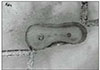
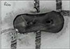
Fig. 1
Pre-instrumented canal at coronal part; Left; Group 1 (SS K file) Right; Group 3 (GT rotary file)
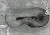
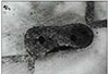
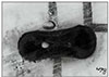
Fig. 2
Post-instrumented canal at coronal part; Left; Group 1 (SS K file) Right; Group 3 (GT rotary file)
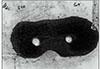
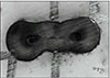
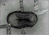
Fig. 3
Pre-instrumented canal at middle part; Left; Group 1 (SS K file) Right; Group 3 (GT rotary file)
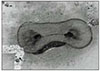
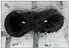
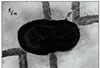
Fig. 4
Post-instrumented canal at middle part; Left; Group 1 (SS K file) Right; Group 3 (GT rotary file)
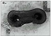
Fig. 5
Pre-instrumented canal at apical part; Left; Group 1 (SS K file) Right; Group 3 (GT rotary file)


References
1. Weine F, Kelly R, Lio P. The effect of preparation procedures on the original canal shape and on apical foramen shape. J Endod. 1975. 1:255–262.

2. Schilder H. Cleaning and shaping the root canal. Dent Clin North Am. 1974. 18:269–296.
3. Weine F. Endodontic therapy. 1989. St. Louis: CV Mosby;277.
4. Walton R. Current concepts of canal preparation. Dent Clin North Am. 1992. 32:309–326.
5. Morgan LA, Montgomery S. An evaluation of the crown-down pressureless technique. J Endod. 1984. 10:491–498.
6. Goerig A, Michelich R, Schultz H. Instrumentation of root canals in molars using the step-down technique. J Endod. 1982. 8:550–554.
7. Roane J, Sabala C, Duncanson M. The balanced force concept for instrumentation of curved canals. J Endod. 1985. 11:203–211.
8. Chenail BL, Teplitsky PE. Endosonics in curved root canals. J Endod. 1985. 11:369–374.
9. Walsh C, ElDeeb ME, Messee HH. Effect of varying ultrasonic power on instrumentation time and prepared canal space. J Endod. 1987. 13:133–138.
10. Walia H, Brantley WA, Gerstein H. An investigation of the bending and torsional properties of Nitinol root canal files. J Endod. 1988. 14:346–351.
11. Esposito PT, Cunningham CJ. A comparison of canal preparation with Nickel-Titanium and Stainless steel instruments. J Endod. 1995. 21:173–176.
12. Baek SH. A Comparison Of Canal Preparation With Engine-Driven Nickel-Titanium Files, Ultrasonic Files, And Stainless Steel Files. J Korean Dent Assoc. 1995. 34(5):363–371.
13. Park HS, et al. A Study On The Shape Of A Canal Prepared With Profiles In A Curved Canal. J Korean Acad Conserv Dent. 1999. 24:633–637.
14. Hata G, Uemura M, Toda T, Kato AS, Imura . A comparison of shaping ability using ProFile, GT file, and Flex-R endodontic instruments in simulated canals. J Endod. 2002. 28(4):316–321.
15. Bramante CM, Berbert A, Borges RP. A Methdology for evaluation of root canal instrumentation. J Endod. 1987. 13:243–245.
16. Calhoun G, Mongomery S. The effects of four instrumentation technique on root canal shape. J Endod. 1988. 14:273–277.
17. Lee SJ, Shin YG, Hwang HK. Transportation of curved canal after canal enlargement according to filing instruments. J Korean Acad Conserv Dent. 1999. 24:503–510.
18. Haga CS. Microscope measurements of root canal preparations folowing instrumentation. J Br Endod Soc. 1968. 2:41–46.
19. Walton RE. Histologic evaluation of different methods of enlarging the pulp canal space. J Endod. 1976. 10:304–310.
20. Mizrahi SJ, Yucker JW, Seltzer S. A scanning electron microscopic study of the efficacy of various endodontic instruments. J Endod. 1975. 1:324–333.
21. Bolanos OR, Jensen JR. Scanning electron microscope comparison of the efficacy of various methods of root cnal preparation. J Endod. 1980. 6:815–821.
22. Tachibana H, Matsumoto K. Applicability of x-ray computerized tomography in endodontics. Endod Dent Traumatol. 1990. 6:16–20.
23. Gambill JM, Alder M, Rio CE. Comparioson of Nickel-Titanium and stainless steel hand-file instrumentation using computed tomography. J Endod. 1996. 22:369–375.
24. Buchanan LS. The shape of things to come. Dentistry today. 1994. 13.
25. Tucker DM, Wenckus CS, Bentkover SK. Canal wall planning by engine-driven Nickel-Titanium instruments, compared with stainless steel hand instrumentation. J Endod. 1997. 23:170–173.
26. Luiten DJ, Morgan LA, Baumgartner JC. A comparison of four instrumentation techniques on apical canal transportation. J Endod. 1995. 21:26–32.
27. Lee JH. The Effect Of Some Canal Preparation Techniques On The Shape Of Root Canals. J Korean Acad Conserv Dent. 1999. 24:337–345.
28. Kosa DA, Marshall G, Baumgartner JC. An analysis of canal centring using mechanical instrumentation techniques. J Endod. 1999. 25:441. 445.
29. Lim KA, Yoon SH. A comparision of stainless steel Kfile, Profile.04, and Quantec LX instruments to shape curved root canals in vitro. J Korean Acad Conserv Dent. 2000. 25:133–140.
30. Glosson CR, Haller RH, Dove SB, Rio CE. A comparison of root canal preparations using NiTi hand, NiTi engine-driven, and K-Flex endodontic instruments. J Endod. 1995. 21:146–151.
31. Baumgartner JC, Martin H, Sabala CL, Strittmatter EJ, Wildey WL, Quigley NC. Histomorphic comparison of canals prepared by four techniques. J Endod. 1992. 17:530–534.
32. Bou Dagher FE, Yared GM. Comparison of three files to prepare curved root canals. J Endod. 1995. 21:264–265.
 XML Download